Real-time X-ray diffraction investigation of metal deformation
- PDF / 1,971,674 Bytes
- 10 Pages / 590.28 x 785 pts Page_size
- 51 Downloads / 283 Views
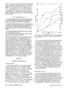
by Green t4j and in 1984 by Winter and Green tS] which concentrated on the direct display of X-ray topographs. Figure 1 shows block diagrams of the two generic electro-optical methods used to permit rapid viewing and recording of X-ray diffraction images. The first is the direct method, which uses a rotating anode, flash generator, or synchrotron radiation as a high-intensity X-ray source. From such a high-intensity source, one obtains a high intensity diffraction image, permitting use of a low gain, high resolution electro-optical imaging system. As also shown in Figure 1, the second is the indirect method, which uses a conventional X - r a y tube source. With this method, a lower intensity source leads to a low intensity diffraction image, which therefore requires a high gain electro-optical imaging system. A variety of combinations can be used, but all indirect systems share the disadvantage that the spatial resolution of the image is limited by the fluorescent screen used to convert X-ray photons into visible light photons. However, all indirect systems have the advantage that they are the least susceptible to radiation damage, permit upgrading with minimum disruption as new components become available, and are the least expensive system design. Two image intensifier tube types which have been most often used to view X-ray diffraction images are shown schematically in Figure 2. By cascading three individual first generation image tube stages shown in Figure 2(a), light gains as high as several million can be obtained. The second generation microchannel plate image intensifier tube shown in Figure 2(b) is similar to a single stage first generation device except for the extremely important addition of a microchannel plate. Since the gain of this type image intensifier tube is several hundred thousand, X-ray diffraction patterns can be successfully viewed in real-time with a single stage when using a synchrotron or flash X-ray source. The visible image from the output of these tubes can be viewed directly, recorded on film, or displayed on a closed circuit television monitor and videotaped. A microchannel plate X-ray detector optimally suited for detection of synchrotron topographic images t61 has been installed on topographic beam line X-19C of the National Synchrotron Light Source at Brookhaven National Laboratory. Recently a new class of cameras has been developed, the solid state silicon devices, which permit direct conversion of photon induced charge distributions to video VOLUME 20A, APRIL 1989--595
HIGH INTENSITY X-RAY SOURCE
/
X-RAY
ELECTRON
FIBEROPTIC
CONVENTIONAL X - RAY GENERATOR
STRONG
VIGIGLEIMAGE
I
HIGH INTENSITY X- RAY BEAM
MONOCHROMI / OR ZAND ING
LOW INTENSITY X-RAY BEAM
i
COLLIMATING SYSTEM
I
I HIGH INTENSITY X-RAY DIFFRACTION IMAGE LOW GAIN X-RAY SENSI T IVE TV CAMERA
MONOCHROMIZING AND / OR COLLIMATING SYSTEM
/
X-RAY
ELECTRON
SCINTILLATOR PHOTO;THODE / ~,~ WEA~~
l
.'~h
PHOSPHOR ST.ONG
I
HIGH INTENSITY X-RAY BEAM
CRYSTAL SPECIMEN
I
PORTABLEIMAGEX-RAYINTENSIFIER (a)
Data Loading...