Role of actin cytoskeleton in podocytes
- PDF / 507,275 Bytes
- 8 Pages / 595.276 x 790.866 pts Page_size
- 43 Downloads / 316 Views
REVIEW
Role of actin cytoskeleton in podocytes Sanja Sever 1 Received: 7 August 2020 / Revised: 14 September 2020 / Accepted: 5 October 2020 # IPNA 2020
Abstract The selectivity of the glomerular filter is established by physical, chemical, and signaling interplay among its three core constituents: glomerular endothelial cells, the glomerular basement membrane, and podocytes. Functional impairment or injury of any of these three components can lead to proteinuria. Podocytes are injured in many forms of human and experimental glomerular disease, including minimal change disease, focal segmental glomerulosclerosis, and diabetes mellitus. One of the earliest signs of podocyte injury is loss of their distinct structure, which is driven by dysregulated dynamics of the actin cytoskeleton. The status of the actin cytoskeleton in podocytes depends on a set of actin binding proteins, nucleators and inhibitors of actin polymerization, and regulatory GTPases. Mutations that alter protein function in each category have been implicated in glomerular diseases in humans and animal models. In addition, a growing body of studies suggest that pharmacological modifications of the actin cytoskeleton have the potential to become novel therapeutics for podocyte-dependent chronic kidney diseases. This review presents an overview of the essential proteins that establish actin cytoskeleton in podocytes and studies demonstrating the feasibility of drugging actin cytoskeleton in kidney diseases. Keywords Glomerular diseases . Podocytes . Actin cytoskeleton . Regulatory GTPases . Actin binding proteins (ABPs) . Nucleators of actin polymerization
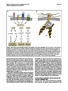
Introduction Podocytes are terminally differentiated epithelial cells of the glomerulus which develop a characteristic architecture specialized for glomerular ultrafiltration. Their structure is traditionally divided into three distinct subcellular compartments: the cell body, primary processes which are microtubule-driven membrane extensions, and foot processes (FPs) which are actin-driven membrane extensions [1]. Adjacent podocytes interdigitate with each other at their FPs which are bridged with a specialized intercellular junction called a slit diaphragm (SD). FPs serve as an adhesive apparatus to the glomerular basement membrane (GBM), which together with endothelial cells and their glycocalyx form a filtration barrier. Regardless of the underlying glomerular disease, the earliest signs of podocyte injury are characterized by the reorganization of FPs structure resulting in a fusion of filtration slits termed “FP effacement” [2–7]. Indeed, for over 50 years, the * Sanja Sever [email protected] 1
Harvard Medical School and Massachusetts General Hospital, Boston, MA, USA
effacement of FPs has been a main feature of proteinuria, though its significance with regard to proteinuria is still a mystery [8]. Recently, a study by Benzing and colleagues suggested that permeability of the renal filter is modulated through compression of the capillary wall [9]. By using CRISPR-Cas9based genome editi
Data Loading...