Self-Affine Fractal Characterization of a TNT Fracture Surface
- PDF / 680,648 Bytes
- 5 Pages / 417.6 x 639 pts Page_size
- 78 Downloads / 294 Views
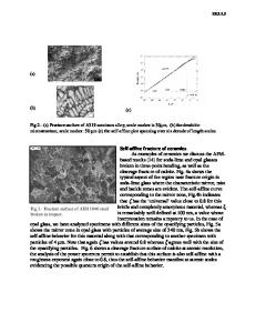
ABSTRACT A trinitrotoluene (TNT) fracture surface image is characterized in terms of a self-affine fractal structure. The fracture surface was produced by high acceleration in an ultracentrifuge when the TNT strength was exceeded. An atomic force microscope (AFM) captured the topography of a 4 micron square region on the fracture surface. The present analysis supports a self-affine description of the TNT fracture surface (wavelengths of 0.016 micron to 4.0 micron) and provides a new prespective on fracture processes in TNT. An essential step in self-affine fractal characterization of surfaces is the determination of reference surfaces. A self-affine fracture surface can be described in terms of a single-valued height function. In the TNT fracture surface, single-valued height functions, which describe surface texture can only be defined with respect to curved reference surfaces. By employing curved reference surfaces, we have demonstrated that self-affine fractal scaling can be used to characterize the TNT fracture surface. This provides important information that is not evident in the analysis of individual surface scans. INTRODUCTION Energetic materials, including details of the rapid chemical reactions that lead to explosive behavior, are of significant interest for scientific and practical reasons. Nevertheless, the details are still poorly characterized and understood. Energetic materials have numerous applications in propulsion systems, in ordnance, and in mining. Because of the conditions under which they are often employed, the nature of fracture processes in such materials under high accelerations is of particular interest. The characterization of fracture in trinitrotoluene (TNT) and in other energetic materials has been the subject of several investigations 1 -4. TNT FRACTURE SURFACE The original TNT sample was melt-cast in a polycarbonate sleeve. The fracture of the sample was produced by high acceleration in an ultracentrifuge when the strength of the material was exceeded. Fracture occurred for an acceleration of 35,000 g at 25'C. The topography of a 4 pm by 4 pm square region (projected approximately perpendicular to the z-axis of the polycarbonate sleeve) square region on the fracture surface was captured by atomic force microscope (AFM). The central 2 pm by 2 pim region (projected) was found to have been deformed by an earlier application of the AFM. The stylus force applied on the previous scan was approximately 12 nanoNewtons, and apparently it was sufficient to produce a permanent compression of the fracture surface. A surface plot of the AFM elevation data (which is composed of 512 uniformly spaced 363
Mat. Res. Soc. Symp. Proc. Vol. 578 ©2000 Materials Research Society
profiles, each of which is composed of 512 uniformly spaced values) is shown in Fig. 1. The "line" visible toward the left hand side of the image was produced by omitting profiles 204 through 216 from the surface plot. This does not affect the analysis. The x-values and y-values correspond to pixel spacings (4 jim/S 12 =0.007
Data Loading...