Ultrasound evaluation of varicoceles: systematic literature review and rationale of the ESUR-SPIWG Guidelines and Recomm
- PDF / 1,945,166 Bytes
- 21 Pages / 595.276 x 790.866 pts Page_size
- 5 Downloads / 304 Views
REVIEW PAPER
Ultrasound evaluation of varicoceles: systematic literature review and rationale of the ESUR‑SPIWG Guidelines and Recommendations Michele Bertolotto1 · Simon Freeman2 · Jonathan Richenberg3 · Jane Belfield4 · Vikram Dogra5 · Dean Y. Huang6 · Francesco Lotti7 · Karolina Markiet8 · Olivera Nikolic9 · Subramaniyan Ramanathan10 · Parvati Ramchandani11 · Laurence Rocher12,13 · Mustafa Secil14 · Paul S. Sidhu6 · Katarzyna Skrobisz8 · Michal Studniarek8 · Athina Tsili15 · Ahmet Tuncay Turgut16 · Pietro Pavlica17 · Lorenzo E. Derchi18 · Members of the ESUR-SPIWG WG Received: 4 June 2020 / Accepted: 11 July 2020 © The Author(s) 2020
Abstract Although often asymptomatic and detected incidentally, varicocele is a relatively common problem in patients who seek medical attention for infertility problems. Ultrasound (US) is the imaging modality of choice for evaluation, but there is no consensus on the diagnostic criteria, classification, and examination technique. In view of this uncertainty, the Scrotal and Penile Imaging Working Group of the European Society of Urogenital Radiology (ESUR-SPIWG) undertook a systematic review of the available literature on this topic, to use as the basis for evidence-based guidelines and recommendations. This paper provides the results of the systematic review on which guidelines were constructed. Keywords Varicocele · Infertility · US · Doppler studies
Introduction Varicocele is defined as dilation of the pampiniform venous plexus draining the testicle, with reflux of venous blood [1, 2]. Although it can be asymptomatic and detected incidentally, it is a relatively common problem in patients who seek medical attention for infertility problems, or complain of chronic scrotal pain or discomfort [3]. Varicocele is detected and graded clinically using the criteria introduced by Dubin and Amelar in 1970, a subjective evaluation which is highly dependent on the expertise of the physician [4]. Colour Doppler ultrasound (US) is the imaging modality of choice [5], but the need for imaging itself is debated. In Europe, the use of US is recommended to confirm clinically The members of “the ESUR-SPIWG WG" are listed in Acknowledgement section. * Michele Bertolotto [email protected] 1
Department of Radiology, University of Trieste, Ospedale Di Cattinara, Strada di Fiume 447, 34149 Trieste, Italy
University Hospitals Plymouth NHS Trust, Derriford Hospital, Derriford Road, Crownhill, Plymouth PL6 8DH, Devon, UK
2
suspected varicoceles, whilst in the USA and in Asia, routine use of imaging is not recommended [5]. Moreover, there is no agreement on how to perform the US examination. A variety of classifications is used based on different sonographic parameters, even within the same country, depending on the practice of the individual sonologist and of the referring clinician [2]. There is a large, but extremely heterogeneous body of published investigations on US imaging of varicoceles. Even though variability makes it impossible to perform a metanalysis, a systematic literature re
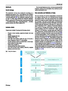
Data Loading...