Selective-Area Growth of Metal Oxide Films Induced by Patterned Excimer Laser Surface Photolysis
- PDF / 1,805,027 Bytes
- 6 Pages / 420.48 x 639 pts Page_size
- 42 Downloads / 347 Views
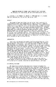
SELECTIVE-AREA GROWTH OF METAL OXIDE FILMS INDUCED BY PATTERNED EXCIMER LASER SURFACE PHOTOLYSIS R. R. KUNZ, D. J. EHRLICH, J. MELNGAILIS, and M. W. HORN Lincoln Laboratory, Massachusetts Institute of Technology, Lexington, Massachusetts 02173 ABSTRACT An excimer-laser-based method for achieving selective-area oxide growth on polymer surfaces is described. This method uses high photon energy (6.4 eV, 7.9 eV) excimer laser irradiation to change hydrophobic surfaces (surface tension 200 erg/cm ) surface. To ensure control of the thickness, the gases pressures are cycled during the growth. As a result, the net growth is not linear with time. If the growth were linear with time, the x-axis used for this figure would be growth time and the figure would be similar to the one shown.
(a)
--
1 Pm
-- I
Co
60
z 40 20
20
40
60
80
THICKNESS GROWN ON HIGH ENERGY SURFACE (nm)
(b)
I•1
100 nm 2
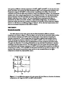
Fig. 6. Surface morphology of SiO 2 grown from SiCI4 and H2 0 on a 15 erg/cm surface at a temperature
of 13 *C. The total growth time was 20 minutes (compared to 2.5 minutes for the film shown in Figure 7), a time long enough for the nuclei to have coalesced into a continuous film.
109
physisorbed layers. The growth on a hydrophilic surface has been studied in some detail in Reference 12, and is correlated to the development of specific adsorbed H2 0 phases. On a hydrophobic surface, where multiple adlayer formation does not occur, the onset of growth occurs only after nucleation of molecular H2 0 clusters. For patterning, the area selectivity of the surface modification process can be estimated from the simple thermodynamics of H 2 0 nucleation. Figure 5 shows relative thickness for growth on hydrophobic surfaces compared to that on a hydrophilic surface. Note the induction time which is characteristic of delayed condensation and results from a thermodynamic nucleation barrier [141. This is further supported by Figure 6, which shows an electron micrograph of SiO 2 grown on a surface whose critical surface tension was 15 erg/cm 2. Note the clearly visible nuclei. In contrast, growth of SiO 2 on high surface energy surfaces results in conformal growth [12]. Understanding the H2 0 nucleation behavior will allow prediction of the ultimate growth selectivities. The H20 nucleation rate I is given by [141 I = k J sin 0 (Gnuc)1/ 2 exp[(Gdes- Gdiff- Gnuc)/kT] where k is a constant, J is the molecular flux to the surface, 0 is the contact angle, Gdes is the activation energy for H20 desorption, Gdiff is the activation energy for H 20 surface diffusion, and Gnuc is the free energy change upon nucleation of a critically sized H 20 cluster. Changes in critical surface tension upon irradiation change 0, Gds, Gdiff, and Gnuc. We have quantitatively calculated Gnuc using cluster radii derived from molecular dynamics calculations [151 and have found that it is only weakly dependent on surface energy, and can be assumed constant.Gdiff and Gdes are coupled and as a simplest estimate, Gdiff can be assumed equal to Gdes/ 2 , so the two terms
Data Loading...