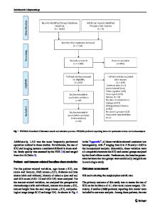
The application of laparoscopy combined with indocyanine green fluorescence imaging technique for hepatic cystic echinoc
- PDF / 1,091,570 Bytes
- 8 Pages / 595.276 x 790.866 pts Page_size
- 94 Downloads / 326 Views
Open Access
RESEARCH ARTICLE
The application of laparoscopy combined with indocyanine green fluorescence imaging technique for hepatic cystic echinococcosis Yu‑Peng Li1, Zhi‑Gang Ma1, Tuerhongjiang Tuxun2, Zhi‑De Li1, Yuan Meng1 and Xiong Chen1*
Abstract Background: With the mature application of laparoscopy in hepatobiliary surgery, laparoscopic treatment of hepatic cystic echinococcosis (CE) has made certain progress. But, due to the inherent limitations of laparoscopy and the growth characteristics of cystic echinococcosis, distinguishing the boundary between cystic lesion and normal hepatic parenchyma is pivotal importance for successful surgery. Indocyanine green (ICG) fluorescence imaging technology can view the boundary of lesion and normal tissue during the treatment of hepatic cystic echinococcosis. Applied laparoscopy combined with ICG fluorescence imaging technique for hepatic cystic echinococcosis may be an effective surgical strategy. Methods: The clinical data contained nine patients with hepatic cystic echinococcosis who underwent laparoscopic surgery with indocyanine green fluorescence imaging technique in authors’ institution from December 2018 to December 2019 were retrospectively analyzed. Indocyanine green was administered intravenously three days prior to surgery. The fluorescence acquisition system for real-time imaging was used during the surgery and the patients were followed up after surgery. Results: Of reported nine patients, six are male and the remaining three are female. The average age is (36.4 ± 7.6) years. For all subjects, surgical procedures were performed under laparoscopy with indocyanine green fluorescence system. This technique showed the clear boundary of the hepatic cyst with normal liver parenchyma. Total cystec‑ tomy in six patients, subtotal cystectomy in two patients and partial hepatectomy in one patient were performed respectively. The average operation time was 3.8 ± 0.9 h, blood loss 206.0 ± 120.7 ml. Neither blood transfusion nor post-operative complication was experienced. The average abdominal drainage time was 3.4 ± 0.9 days with hospital stay 5.7 ± 2.1 days. During the 6–12 months follow-up period, neither recurrence nor intraperitoneal implantation was found. Conclusions: Applied laparoscopy combined with ICG fluorescence imaging technique for hepatic cystic echinococ‑ cosis is safe and feasible. Enhanced boundary image can assist surgeons to complete radical resection and reduce complications. Keywords: Hepatic cystic echinococcosis, Laparoscopy, Pericystectomy, Indocyanine green (ICG), Fluorescence imaging, Boundary
*Correspondence: [email protected] 1 Department of Hepatobiliary Surgery, People’s Hospital of Xinjiang Uygur Autonomous Region, No. 91 Tianchi Road, Urumqi 830000, Xinjiang Uygur, China Full list of author information is available at the end of the article
Background Hepatic cystic echinococcosis is a zoonosis caused by the larval stage of Echinococcus granulosus and continues to be a public health threat in northwest China. Despite the
Data Loading...