The histopathological evaluation based on the indocyanine green fluorescence imaging of regional lymph node metastasis o
- PDF / 824,823 Bytes
- 7 Pages / 595.276 x 790.866 pts Page_size
- 64 Downloads / 309 Views
ORIGINAL ARTICLE
The histopathological evaluation based on the indocyanine green fluorescence imaging of regional lymph node metastasis of splenic flexural colon cancer by near-infrared observation Manabu Kakizoe 1 & Jun Watanabe 2 & Yusuke Suwa 2 & Kazuya Nakagawa 3 & Hirokazu Suwa 1 & Mayumi Ozawa 3 & Atsushi Ishibe 3 & Hidenobu Masui 1 & Kaoru Nagahori 1 Accepted: 10 November 2020 # Springer-Verlag GmbH Germany, part of Springer Nature 2020
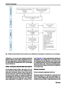
Abstract Purpose The purpose of this study was to investigate the relationship between the fluorescence on indocyanine green fluorescent imaging (ICG-FI) and the histopathological findings of regional lymph node (LN) metastasis of splenic flexural colon cancer. Methods From July 2013 to December 2018, consecutive patients with splenic flexural colon cancer with a preoperative diagnosis of N0 who underwent laparoscopic surgery were enrolled. The distribution of cancer sites in metastatic LNs (completely/not completely occupied by metastatic foci) was evaluated with hematoxylin and eosin–stained preparations. We compared the relationship between the distribution of cancer site and fluorescence of paraffin block in metastatic LNs. Results Seventy-two patients were enrolled, of whom 13 (18.1%) had metastatic LNs. A total of 25 metastatic LNs were evaluated. The median short axis of the occupied LNs was 4.5 mm, which was significantly larger than that of the nonoccupied LNs (3.0 mm; p = 0.036). In the near-infrared observation of the paraffin block, the completely occupied LNs showed no fluorescence, regardless of the LN size, but 8 of 10 non-occupied LNs showed fluorescence (p < 0.001). Even the nonoccupied LNs that showed fluorescence, the cancer site did not show fluorescence. Conclusions The occupied LNs showed no fluorescence, but 80% of the non-occupied LNs showed fluorescence. Even in nonoccupied LNs that showed fluorescence, the cancer site did not show fluorescence. This demonstrated LN dissection should not be omitted, even if no fluorescence is noted on intraoperative ICG-FI. Keywords Colorectal cancer . Lymph node metastasis . Indocyanine green . Near infrared . Fluorescence
Introduction In the last two decades, the usefulness of intraoperative navigation surgery has been reported in various surgical fields
* Jun Watanabe [email protected] 1
Department of Surgery, Yokosuka Kyosai Hospital, Yokosuka, Japan
2
Department of Surgery, Gastroenterological Center, Yokohama City University Medical Center, 4-57, Urafune-cho, Minami-ku, Yokohama 232-0024, Japan
3
Department of Gastroenterological Surgery, Yokohama City University Graduate School of Medicine, Yokohama, Japan
[1–7]. In particular, indocyanine green (ICG) fluorescence imaging (FI) has been used in clinical practice for colorectal cancer surgery to evaluate the lymph flow for optimal lymph node (LN) dissection and anastomotic perfusion in the field of colorectal cancer surgery [8–14]. We previously reported that the lymph flow evaluation for colon cancer located in the splenic flexure (SFx) using IC
Data Loading...