Ultra-fast High Resolution Microscopy: Options for Pump-probe Methods
- PDF / 171,488 Bytes
- 12 Pages / 612 x 792 pts (letter) Page_size
- 61 Downloads / 345 Views
1026-C12-01
Ultra-fast High Resolution Microscopy: Options for Pump-probe Methods Archie Howie Physics, University of Cambridge, Madingley Road, Cambridge, CB3 0HE, United Kingdom ABSTRACT Recent applications of pulsed pump-probe techniques in microscopy are described, often going hand in hand with improved spectroscopy. These methods can provide information about dynamic response extending down towards the femto-second (fs) timescale for re-settable, repeatable phenomena and may thus help to fill a large gap in the microscopist’s coverage of material behavior. Although the use of photon pulses is best developed, ultra-short electron pulses are also now available. Various options are therefore to hand in either transmission electron microscopy or scanning transmission microscopy operation for combining the high spatial resolution of electrons with the spectral selectivity and precision of photons. With purely photon pump-probe operation, good spatial resolution can perhaps be obtained using the tip field-enhancement effect in scanning probe microscopy (SPM) and has also recently been impressively demonstrated in two-photon photo-electron microscopy (PEEM) operation. INTRODUCTION Electron microscopy now provides impressive static structural information, at least in two-dimensional projection, over a wide range extending down to the 0.1nm level. In the frequency – time domain of spectroscopy or dynamic imaging however, an enormous gap still exists [1] between the current lower limit of about 1eV in electron energy loss spectroscopy (EELS), equivalent to about 4 fs in time, and the upper 30 Hz frequency of high resolution electron microscopy (HREM) video recording. This gap is depicted in fig. 1 and closing of it by improvements either in spectral range and accuracy or in dynamic microscopy can bring benefits to both the time and frequency domains. In special cases, such as the thermal motion of the atoms in a perfect crystal, (q,ω) spectroscopy can provide a complete description and it is possible to establish the most important details even more simply from neutron powder diffraction data [2]. Of course spectroscopy and dynamic imaging are not in general fully equivalent since the Fourier relation between them is similar to that between imaging and diffraction and applies to complex amplitudes not to measured intensities. This point is illustrated by the familiar recipe for the time-dependence of the wave function ψ(r,t) of a quantum mechanical system.
ψ(r,t) = Σn an φn(r) exp(-iωnt)
(1)
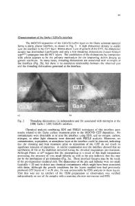
Although it does not give information about the phases of either the wave amplitudes an or eigen-functions φn(r), spatially localized spectroscopy yields the energy eigenvalues ħωn, eigenmode intensities │φn(r)│2 and perhaps also the occupation levels │an│2. Even without the benefits of dynamic microscopy, there is therefore useful mileage in extending the range of spatially resolved spectroscopy. These possibilities are well exemplified by the use of the cathodoluminescence (CDL) signal to display the different surface plasmon
Data Loading...