Widespread cortical dyslamination in epilepsy patients with malformations of cortical development
- PDF / 7,836,660 Bytes
- 10 Pages / 595.276 x 790.866 pts Page_size
- 56 Downloads / 269 Views
FUNCTIONAL NEURORADIOLOGY
Widespread cortical dyslamination in epilepsy patients with malformations of cortical development Eyal Lotan 1,2,3 & Omri Tomer 4 & Ido Tavor 1,2 & Ilan Blatt 2,5 & Hadassah Goldberg-Stern 2,6 & Chen Hoffmann 1,2 & Galia Tsarfaty 1,2 & David Tanne 2,7 & Yaniv Assaf 4,8 Received: 11 June 2020 / Accepted: 16 September 2020 # Springer-Verlag GmbH Germany, part of Springer Nature 2020
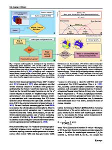
Abstract Purpose Recent research in epilepsy patients confirms our understanding of epilepsy as a network disorder with widespread cortical compromise. Here, we aimed to investigate the neocortical laminar architecture in patients with focal cortical dysplasia (FCD) and periventricular nodular heterotopia (PNH) using clinically feasible 3 T MRI. Methods Eighteen epilepsy patients (FCD and PNH groups; n = 9 each) and age-matched healthy controls (n = 9) underwent T1 relaxation 3 T MRI, from which component probability T1 maps were utilized to extract sub-voxel composition of 6 T1 cortical layers. Seventy-eight cortical areas of the automated anatomical labeling atlas were divided into 1000 equal-volume sub-areas for better detection of cortical abnormalities, and logistic regressions were performed to compare FCD/PNH patients with healthy controls with the T1 layers composing each sub-area as regressors. Statistical significance (p < 0.05) was determined by a likelihood-ratio test with correction for false discovery rate using Benjamini-Hochberg method. Results Widespread cortical abnormalities were observed in the patient groups. Out of 1000 sub-areas, 291 and 256 bilateral hemispheric cortical sub-areas were found to predict FCD and PNH, respectively. For each of these sub-areas, we were able to identify the T1 layer, which contributed the most to the prediction. Conclusion Our results reveal widespread cortical abnormalities in epilepsy patients with FCD and PNH, which may have a role in epileptogenesis, and likely related to recent studies showing widespread structural (e.g., cortical thinning) and diffusion abnormalities in various human epilepsy populations. Our study provides quantitative information of cortical laminar architecture in epilepsy patients that can be further targeted for study in functional and neuropathological studies. Keywords MRI . Cortical layers . T1 relaxation . Focal cortical dysplasia . Periventricular nodular heterotopia . Epilepsy
DT and YA contributed equally to this work. * Eyal Lotan [email protected] 1
Department of Diagnostic Imaging, Sheba Medical Center, Tel Hashomer, 52621 Ramat Gan, Israel
2
Sackler Faculty of Medicine, Tel Aviv University, 69978 Tel Aviv, Israel
3
Department of Radiology, NYU Langone Medical Center, 660 1st Ave, New York, NY 10016, USA
4
Sagol School of Neuroscience, Tel Aviv University, 69978 Tel Aviv, Israel
5
Department of Neurology, Sheba Medical Center, Tel Hashomer, 52621 Ramat Gan, Israel
6
Department of Neurology, Schneider Children’s Medical Center of Israel, 49202 Petah Tikva, Israel
7
Stroke Center, Department of Neurology and Sa
Data Loading...