Dose and Doping Dependence of Damage Annealing in Fe Mev Implanted Inp
- PDF / 3,006,473 Bytes
- 6 Pages / 414.72 x 648 pts Page_size
- 70 Downloads / 416 Views
becomes crucial to understand the complex processes that may occur during post-implantation annealing treatments. In fact, Fe is known to strongly react with implantation damage. Moreover, preliminary investigations [9] indicated the role of amorphous layer recovery when the implanted dose exceeds the amorphization threshold: gettering and/or precipitation of Fe was observed in connection with the solid phase epitaxial growth of the amorphised region. In the present paper we report on progress in characterising room temperature Fe MeV implantation and the subsequent annealing process. The role of implanted dose, initial doping of the substrates, and of the annealing temperature is examined and the relation between the Fe redistribution and the defect structure is discussed. A comparison between RT implantation and the results of high implantation temperature experiments (200 'C) is also presented and discussed. EXPERIMENT Room temperature (RT) Fe implantation was performed at 2 MeV energy on (100) InP wafers, 3 both Sn doped (1.5x10 18 cm-3 ) and undoped (slightly n-type, < IxI0 16 cm- ). The implanted 2 doses ranged from 5xI0 13 to 2x10 14 at/cm 2, while a current density of the order of 10 nA/cm was the and in this case performed °C) were also implants (200 temperature Some elevated employed. doses ranged from 2x10 14 to 5x10 14 at/cm 2. The implanted samples were annealed at temperatures ranging from 650 'C to 800 'C for times varying between 1 and 1.5 hours. The chamber of a MOCVD reactor was employed to mantain a phosphorous overpressure in the annealing ambient, so as to avoid surface decomposition due to P evaporation. 829 Mat. Res. Soc. Symp. Proc. Vol. 396 ©1996 Materials Research Society
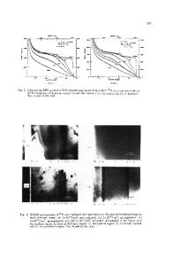
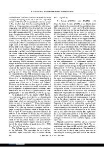
The structure of the damage produced by the implantation process and its evolution with annealing were studied by means of RBS-Channeling and TEM. RBS spectra were recorded after impinging the samples with a 3.5 MeV 4 He+ beam aligned with the crystal axis and detecting the backscattered ions at a 170' angle with respect to the beam direction. Cross sectional TEM analyses were performed in both bright/dark field and High Resolution modes, employing a JEOL 2000FX instrument. Selected area and micro-diffraction patterns were also recorded to assess the degree of amorphization of the as-implanted samples. Thinning of the samples was performed by Ar ion milling at the liquid nitrogen temperature in order to avoid any modification of the damage structure due to heating up of the samples (this is particularly important for as implanted samples). Fe depth profiles were obtained by SIMS, using a CAMECA IMS4f instrument. A 8 keV 02+ primary beam was employed and the 56Fe+ secondary ions were detected. The sensistivity factors
for quantifying the profiles were obtained by measuring an implanted standard of known dose under the same experimental conditions.
RESULTS In fig. 1 the results of the RBS-Channeling measurements for the 5x10 13 and 2x10 14 cm-2 RT implanted samples (undoped substrate) are compared. The RBS yield of the
Data Loading...