Engineered Nanostructured Coatings for Enhanced Protein Adsorption and Cell Growth
- PDF / 360,568 Bytes
- 7 Pages / 432 x 648 pts Page_size
- 60 Downloads / 385 Views
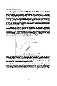
Engineered Nanostructured Coatings for Enhanced Protein Adsorption and Cell Growth Fereydoon Namavar1, Alexander Rubinstein2, Renat F. Sabirianov2, Geoffrey M. Thiele3, J. Graham Sharp4, Utsav Pokharel1, Roxanna M. Namavar1 and Kevin L. Garvin1 1 Department of Orthopaedic Surgery and Rehabilitation, UNMC, Omaha, NE 68198, U.S.A. 2 Department of Physics, University of Nebraska, Omaha, NE 68182, U.S.A. 3 Department of Internal Medicine Rheumatology, UNMC, Omaha, NE 68198, U.S.A. 4 Department of Genetics, Cell Biology and Anatomy, UNMC, Omaha, NE 68198, U.S.A. ABSTRACT We designed and produced pure cubic zirconia (ZrO2) ceramic1 coatings by an ion beam assisted deposition (IBAD) with nanostructures comparable to the size of proteins. Our ceramic coatings exhibit high hardness and a zero contact angle with serum. In contrast to hydroxyapatite (HA), nano-engineered zirconia films possess excellent adhesion to all orthopaedic materials. Cell adhesion and proliferation experiments were performed with a bona fide mesenchymal stromal cell line (OMA-AD). Our experimental results indicate that the nano-engineered cubic zirconia is superior in supporting growth, adhesion, and proliferation. Since cell attachment is mediated by adhesive proteins such as fibronectin (FN), to elucidate why cells attach more effectively to our nanostructures, we performed a comparative analysis of adsorption energies of FN fragment using quantum mechanical calculations and Monte Carlo (MC) simulation both on smooth and nanostructured surfaces. We have found that a FN fragment adsorbs significantly stronger on the nanostructured surface than on the smooth surface2. INTRODUCTION There is a perpetually growing need to develop methods to enhance or prevent cell adhesion and proliferation as needed and to regulate cellular growth in order to improve health and prevent disease. In recent years, this necessity has led to a heightened interest in studying the interaction of living cells with nanomaterials. Increasing biointegration of prosthetic surfaces not only will lead to faster tissue integration and vascularization, but also will result in faster patient recovery and the savings of a substantial amount of healthcare dollars. By mimicking the nanostructure of the lotus leaf and applying ion beam assisted deposition (IBAD), we have demonstrated the fabrication of pure cubic ZrO2 (a diamond simulant) coating with 2-20 nm grain size (see Fig. 1) which is comparable to the protein dimensions. Comparing this engineered coating to orthopaedic grades of CoCrMo and HA3 with a microcrystalline structure indicates that our engineered nanostructures are superior in supporting the adhesion and proliferation of osteoblasts-like cells of a mouse bone marrow stromal cell line. One of the goals of this paper is to understand why mammalian cells attach more effectively to nanostructured ZrO2 surfaces as compared to the conventional orthopaedic materials. These cells sense their environment and respond accordingly. Surface wettability, morphology, and electric charge c
Data Loading...