Flavours and Fragrances
Flavours and fragrances constitute some of the most beautiful and amazing facets of chemistry. Whereas in many respects chemistry is a very purpose-oriented science, perfumes (and to a lesser extent flavours) may be regarded as luxury items which conspicu
- PDF / 9,958,594 Bytes
- 124 Pages / 476.22 x 680.315 pts Page_size
- 41 Downloads / 314 Views
cents The process of smelling begins when scents in the inhaled air approach the olfactory epithelium (Regio olfactoria) (Fig. 3.1 and 3.2). Richard Axel and Linda B. Buck (Nobel Prize laureates for Pharmacy or Medicine in 2004) identified 339 intact, trans-membrane olfactory receptor proteins, to which transport protein-associated scent molecules can bind. In general, scent molecules bind to various receptor proteins, and on each receptor protein bind different scent molecules. Thus, for each scent results a characteristic binding profile. [1] The olfactory receptors are arranged on whip-like scent hairs, held together in bundles of up to ten cilia; these are found on the 20 million olfactory cells. [2] Each of these cells expresses only one scent receptor. By binding of the scent molecule to the receptor, the quaternary structure of the receptor protein is
i Humans use around 1 % of their genome in order to code the scent receptor proteins, which belong to the family of rhodopsin-like G-protein-coupled receptors (GPCRs).
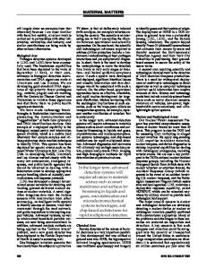
3.1 Cross-section of the human skull (a) and a single olfactory receptor neuron of a frog (Rana pipiens) (b) (c: cilia; d: dendrite; s: perikaryon; a: axonal segment; calibration bar is 10 μm). B. Schaefer, Natural Products in the Chemical Industry, DOI 10.1007/978-3-642-54461-3_3, © Springer-Verlag Berlin Heidelberg 2014
46
3 Flavours and Fragrances
3.2 The smelling process of humans.
3.3 The excitation pattern, which a scent releases in the olfactory bulb, can be visualised in living mice using functional magnetic resonance imaging (fMRI). Shown are the patterns for butanal (C4), pentanal (C5), hexanal (C6), heptanal (C7) and octanal (C8) (high activity, red; slight activity, blue).
a ltered, which leads to a depolarisation of the olfactory membrane and eventually to the collapse of the electrical potential of the cell. This triggers an electrical impulse, which is conducted by olfactory sensory neurons (Filia olfactoria) through the cribriform plate in the upper nasal cavity into the olfactory bulb in the forebrain. There are to be found clusters of neurons called glomeruli, each of which at any one time is assigned to one receptor protein. Each scent produces a characteristic glomeruli-excitation pattern (Fig. 3.3). [3] The excitation pattern represents all olfactory properties of the scent molecule, which are forwarded via the olfactory tract (Tractus olfactorius) into the rhinencephalon and other parts of the brain. The perception is compared with characteristic olfactory impressions in the memory, which finally enables humans to distinguish a diversity of approximately 10,000 scents (Fig. 3.4). [4]
Human Scent The human body has two to four million sweat glands (or sudoriferous glands, from sudor (lat.) sweat), which are devided into two main groups: The eccrine and the apocrine sweat glands. The eccrine sweat glands are distributed almost all over the human body and are utilised for cooling. The apocrine sweat glands
Cilia olfactoria Filia olfactoria Bulbus olfactoriu
Data Loading...