Gelatin Loaded Titanium Dioxide and Silver Oxide Nanoparticles: Implication for Skin Tissue Regeneration
- PDF / 966,001 Bytes
- 12 Pages / 595.276 x 790.866 pts Page_size
- 48 Downloads / 323 Views
Gelatin Loaded Titanium Dioxide and Silver Oxide Nanoparticles: Implication for Skin Tissue Regeneration Nermeen Eldebany 1 & Mohamed Abd Elkodous 2,3 & Hossam Tohamy 4 & Ramadan Abdelwahed 1 & Mahmoud El-kammar 1 & Howaida Abou-Ahmed 1 & Hoda Elkhenany 1 Received: 24 September 2020 / Accepted: 9 November 2020 # Springer Science+Business Media, LLC, part of Springer Nature 2020
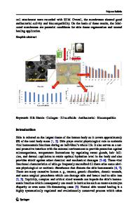
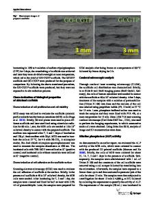
Abstract Treatment of burn wounds has many requirements to ensure wound closure with healthy tissue, increased vascularization, guarantee edema resolution, and control bacterial infection. We propose that titanium oxide (TiO2) nanoparticles (NPs) will be more efficient than silver dioxide (Ag2O) in the treatment of burn wounds. Herein, gelatin loaded NPs (GLT-NPs) were evaluated for their efficacy to regenerate second-degree burn wound in rabbit skin. TEM results revealed that the average particle sizes were 7.5 and 17 nm for Ag2O and TiO2 NPs, respectively. The results of the in vivo application of GLT-NPs on burn wound in the rabbit revealed that both Ag2O and TiO2 NPs were efficient than the control none treated (CTRL) and GLT group. In terms of the healing rate, the GLT-TiO2 did not show any significant difference than GLT-Ag2O (99.57% vs. 99.85%, p = 0.2). Meanwhile, the healing rate was significantly higher in both NPs’ treated groups than CTRL (94.16%, p < 0.01) and GLT group (95.07%, p < 0.05). Also, the histological analysis using H&E staining showed re-epithelization, less edema, and enhanced vascularization in both GLT-NPs than CTRL and GLT groups. Furthermore, immunohistochemical analysis of TGF-β1 and α-SMA revealed significantly a higher expression in both GLT-NPs groups than CTRL and GLT groups at weeks 1 and 2 (p < 0.05). Interestingly, TGF-β1 and α-SMA were substantially higher in GLT- TiO2 than GLT-Ag2O at weeks 1 and 2 (p < 0.05), but the expression was not significant at week 3. In conclusion, GLT-NPs showed higher regenerative capacity and enhanced the healing quality after burn wound compared to CTRL and GLT. Keywords Nanoparticles . Second-degree burn wound . Rabbit . Healing . Vascularization
Introduction Burn wound hazardous is summarized in a couple of points [1]. First, burn injury has a high infection susceptibility due to excessive exudate production, which considers as a suitable microenvironment for bacterial growth [2]. Second, it results in severe tissue degeneration, which leaves a high scar tissue
* Hoda Elkhenany [email protected] 1
Department of Surgery, Faculty of Veterinary Medicine, Alexandria University, Alexandria 21944, Egypt
2
Department of Electrical and Electronic Information Engineering, Toyohashi University of Technology, 1-1 Hibarigaoka, Tempaku-cho, Toyohashi, Aichi 441-8580, Japan
3
Center for Nanotechnology (CNT), School of Engineering and Applied Sciences, Nile University, Giza, Egypt
4
Department of Pathology, Faculty of Veterinary Medicine, Alexandria University, Alexandria 21944, Egypt
depending on the burn degree. Hence, a suitable therapeutic protocol would cover these
Data Loading...