Preparation, Characterization and Optical Properties of Zinc Oxide Nanoparticles
- PDF / 391,602 Bytes
- 6 Pages / 412.92 x 646.2 pts Page_size
- 68 Downloads / 407 Views
OF ZINC OXIDE NANOPARTICLES
SHOUTIAN LI, STUART J. SILVERS and M. SAMY EL-SHALL* Department of Chemistry, Virginia Commonwealth University Richmond, VA 23284-2006
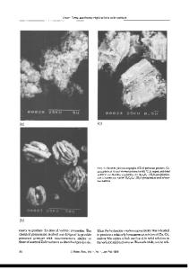
ABSTRACT ZnO nanoparticles are produced by the laser vaporization-controlled condensation technique. These particles are connected in a web-like agglomeration. Their properties are
compared to those of ZnO nanoparticles produced by solution sol-gel and reverse micelle techniques. All particles have the bulk crystal structure and show quantum size effects in
absorption and emission. They show emissions that consist of a blue bandgap feature with a sub-nanosecond lifetime and a green feature with multiexponential lifetime decays. Emission from the stearate coated particles produced by the reversed micelle method is particularly
intense.
INTRODUCTION
Nanoparticles have been the focus of many recent studies because their finite size and
large surface to volume ratio can result in novel properties not exhibited by the bulk material
[1-3]. Such properties are both intrinsically interesting and have important applications in areas such as microelectronics, catalysis, and optical communications. Zinc oxide, a semiconductor, has been prepared as nanoparticles in transparent colloidal solutions by Bahnemann et al. [4], Henglein and co-workers [5,6], Spanhel and Anderson [7] and others. These particles exhibit quantum size effects [8,9]: their bandgap absorption and emission are blue-shifted with respect to bulk ZnO and their visible emission, in the 500 nm region, shows wavelength and lifetime dependence on size. Surface alterations cause noticeable photophysical changes. Here ZnO nanoparticles are prepared by the laser vaporization-controlled condensation
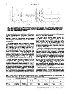
technique (LVCC) and by sol-gel and reversed micelle methods. The properties of these particles, produced by different methods, are compared. EXPERIMENTAL Preparation of ZnO Nanoparticles (1) By the Laser Vaporization-Controlled Condensation Technique. A modified, upward thermal diffusion cloud chamber, shown in Figure 1, is used to prepare ZnO nanoparticles [10-12]. The chamber is filled to 800 Torr total pressure with a known mixture of 02 and He gases. The top plate is cooled to about 150 K with circulating liquid nitrogen. A Zn metal rod (Aldrich) is set on the bottom plate which is maintained at a temperature higher than the upper one (300-400 K). The 532 nm second harmonic of a pulsed Nd:YAG laser is used to evaporate the target. Zn atoms/clusters react with 02 to form ZnO clusters which are carried by convection to the nucleation zone. The particles grow in the nucleation zone and are deposited on the top plate. (2) By the Sol-Gel Method. ZnO nanoparticles in methanol are prepared by the method of Henglein and co-workers [5,6]. A 10 ml sample of 0.1 M Zn(OAc) 2 (Aldrich) in methanol is diluted with 190 ml of methanol. To this is added 4 ml of 10 M NaOH under strong stirring. (3) By a Reversed Micelle Technique. ZnO nanoparticles, coated with stearate, are prepared in a water-in-oil microemulsion
Data Loading...