A deep-red emission fluorescent probe with long wavelength absorption for viscosity detection and live cell imaging
- PDF / 2,819,036 Bytes
- 8 Pages / 595.276 x 790.866 pts Page_size
- 87 Downloads / 279 Views
RESEARCH PAPER
A deep-red emission fluorescent probe with long wavelength absorption for viscosity detection and live cell imaging Li Chen 1 & Yangzhen Feng 1 & Yecheng Dang 1 & Cheng Zhong 2 & Dugang Chen 1 Received: 22 January 2020 / Revised: 7 August 2020 / Accepted: 24 August 2020 # Springer-Verlag GmbH Germany, part of Springer Nature 2020
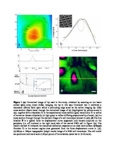
Abstract Intracellular viscosity is closely related to a series of biological processes and could be a biomarker for various diseases. Herein, we reported a deep-red emission viscosity probe ACI, which showed a turn-on fluorescence effect with excellent selectivity encountering high viscous medium. To assure the practical biological application, ACI demonstrated not only a long wavelength emission at 634 nm but also a long wavelength excitation at 566 nm, which were crucial to afford deeper penetration depth and higher sensitivity in bioimaging. The photophysical properties and viscosity recognition mechanism of the probe were carefully discussed here. Theoretical calculations furtherly confirmed that high viscous medium could inhibit the twisted intramolecular charge transfer (TICT) process of the probe which quenched the fluorescence in low viscous media, and restore the emission. More importantly, it was successfully applied to visualize the viscosity in living cells. Keywords Fluorescence . Fluorescent probe . Viscosity . Biosensors . UV/VIS
Introduction Intracellular viscosity is closely related to a series of diffusionmediated biological processes, including mass transportation, chemical signal transporting, metabolite diffusion, and biomacromolecular interaction [1–3]. Abnormal levels of viscosity in living cells could be a valuable biomarker for certain diseases, such as diabetes, Alzheimer’s disease, atherosclerosis, and hypertension [4–6]. Therefore, it is of great importance to quantify the intracellular viscosity which could offer significant information for disease diagnosis and curative effect evaluation. However, conventional techniques for macroscopic fluid viscosity detection using a rotational viscometer, Electronic supplementary material The online version of this article (https://doi.org/10.1007/s00216-020-02911-2) contains supplementary material, which is available to authorized users. * Cheng Zhong [email protected] * Dugang Chen [email protected]; [email protected] 1
Key Laboratory for Green Chemical Process of Ministry of Education, School of Chemical Engineering and Pharmacy, Wuhan Institute of Technology, Wuhan 430205, Hubei, China
2
College of Chemistry and Molecular Science, Wuhan University, Wuhan 430072, Hubei, China
a capillary viscometer, or a falling ball viscometer are totally not proper for microscopic intracellular application [7]. Hence, the development of a new and applicable tool for cellular viscosity measurement is in great demand. Fluorescent probes have showed great advantages and gained much success in the sensing and imaging field of intracellular ions, active small molecules, and enzymes, benefiting from their evident s
Data Loading...