A direct comparison of adenosine and regadenoson myocardial perfusion reserves measured by MRI
- PDF / 285,697 Bytes
- 2 Pages / 610 x 792 pts Page_size
- 3 Downloads / 315 Views
BioMed Central
Open Access
Poster presentation
A direct comparison of adenosine and regadenoson myocardial perfusion reserves measured by MRI Edward DiBella*, Tae Ho Kim, Nathan Pack, Liyong Chen, Henry Buswell, Sirisha Yarlagadda, Alexis Harrison and Sheldon E Litwin Address: University of Utah, Salt Lake City, UT, USA * Corresponding author
from 13th Annual SCMR Scientific Sessions Phoenix, AZ, USA. 21-24 January 2010 Published: 21 January 2010 Journal of Cardiovascular Magnetic Resonance 2010, 12(Suppl 1):P220
doi:10.1186/1532-429X-12-S1-P220
Abstracts of the 13th Annual SCMR Scientific Sessions - 2010
Meeting abstracts - A single PDF containing all abstracts in this Supplement is available here. http://www.biomedcentral.com/content/files/pdf/1532-429X-11-S1-infoThis abstract is available from: http://jcmr-online.com/content/12/S1/P220 © 2010 DiBella et al; licensee BioMed Central Ltd.
Introduction Effective pharmacological vasodilation is essential for assessing myocardial ischemia with MRI. Recently, regadenoson (Lexiscan™) was FDA approved and has been shown to produce comparable SPECT perfusion defects to those seen with adenosine, while causing fewer side effects.
Purpose To directly compare myocardial perfusion reserve with adenosine and regadenoson using quantitative MRI techniques.
the arterial input functions into gadolinium concentration to remove the effects of saturation. Images were registered and segmented to give time curves from 6 tissue regions per slice. The curves were fit to a two compartment model and Ktrans used as an index of perfusion.
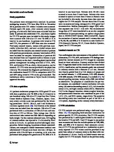
Results Fig. 1 shows the correlation of the perfusion reserves for the two vasodilators. The mean perfusion reserve was 2.34 ± 0.85 for adenosine, and 2.36 ± 1.1 for regadenoson (p = ns). All subjects tolerated both vasodilators well. 6 out of the 8 subjects felt that the regadenoson was easier to tolerate, Figure 1.
Methods 8 subjects (5 female, 3 male) without ischemia were imaged on a 3 T Siemens Trio system. Imaging was done first at rest, and then during adenosine infusion (140 ug/ kg/min) and 34 ± 4 minutes later with regadenoson injection (0.4 mg/5 ml). A 5 cc/sec injection of Gd-BOPTA (Multihance™) was used, with doses of 0.02, 0.03 and 0.03 mmol/kg, respectively. The contrast was injected ~3 minutes after the start of the adenosine infusion, and ~90 seconds after the regadenoson injection. A saturation recovery radial turboFLASH sequence was used with 72 rays acquired after each saturation pulse. Scan parameters were TR = 2.6 msec, TE = 1.14 msec, flip = 14, slice thickness = 8 mm. Reconstruction was performed by iteratively minimizing a cost function as in [1] with total variation constraints in both space and time dimensions. Processing was performed in a manner similar to [2] to convert
Conclusion Regadenoson is a potential alternative to adenosine for use with cardiac MRI studies. Unlike adenosine, regadenoson does not require the use of an MRI-safe infusion pump and the study can be done with one intravenous line rather
Data Loading...