Adherens junctions remain dynamic
- PDF / 1,059,943 Bytes
- 4 Pages / 595 x 794 pts Page_size
- 24 Downloads / 298 Views
Open Access
COMMENTARY
Adherens junctions remain dynamic Commentary Matthias M Falk
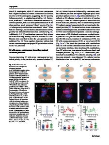
Abstract One of the four principal categories of cell-cell junctions that hold together and shape distinct tissues and organs in vertebrates, adherens junctions (AJs) form cell-cell contacts that connect transmembrane proteins with cytoskeletal actin filaments to provide architectural strength, aid in morphogenesis, and help to maintain proper tissue homeostasis. The classical organization of AJs, consisting of transmembrane cadherins and cytoplasmically attached βcatenins and α-catenins assembled together into a multiprotein complex, was once thought obligatory to craft a robust and stable connection to actin-based cytoskeletal elements, but this architecture has since been challenged and questioned to exist. In a stimulating paper published in a recent issue of BMC Biology, Millán et al. provide convincing evidence that in confluent vascular endothelial cells a novel dynamic vascular endothelial (VE)-cadherinbased AJ type exists that interacts with and physically connects prominent bundles of tension-mediating actin filaments, stress fibers, between neighboring cells. Stress fibers were known previously to link to integrin-based focal adhesion complexes but not to cell-cell adhesion mediating AJs. These new findings, together with previous results support the concept that different AJ subtypes, sharing the same transmembrane cadherin types, can assemble in various configurations to either increase barrier function and promote physical cell-cell adhesion, or to lessen cell-cell adhesion and promote cell separation and migration. Commentary Cells in vertebrates including humans can be linked together by four principal types of cell-cell junctions to form tissues and organs. Each type of cell-cell junction is considered to fulfill a special function. Tight junctions (TJs) form a net-like belt of branched ridges of transmembrane proteins (claudins, occludins, tricellulin) around cells that tightly link cells together to separate apical from basolateral membrane domains, or (in the case of epithelia and vascular endothelia) to separate, outside from inside, or the lumen of blood vessels from the surrounding body, respectively. Desmosomes and adherens junctions (AJs) form patchy cell-cell contacts that connect cytoskeletal elements, such as intermediate filaments and actin filaments of neighboring cells to provide tissue strength, acting as an aid in tissue morphogenesis during development, and to maintain proper tissue organization. Gap junctions (GJs) are clusters of double-membrane-spanning hydrophilic channels made from transmembrane connexin proteins that provide direct cell-to-cell communication by allowing the passage of sig* Correspondence: [email protected] 1
Department of Biology, Lehigh University, Bethlehem, PA, USA
Full list of author information is available at the end of the article
naling molecules, ions, and electrical currents. All four junction types provide physical cell-cell coupling. Epithelia and endothelia,
Data Loading...