Assessment of Geometrical and Transport Properties of a Fibrous C/C Composite Preform Using x-ray Computerized Micro-tom
- PDF / 2,080,877 Bytes
- 12 Pages / 612 x 792 pts (letter) Page_size
- 8 Downloads / 249 Views
nd partially infiltrated carbon–carbon composite preforms have been scanned by high-resolution synchrotron radiation x-ray computerized micro-tomography. Three dimensional high-quality images of the pore space have been produced at two distinct resolutions and have been used for the computation of geometrical quantities: porosity, internal surface area, pore sizes, and their distributions, as well as local and average fiber directions. Determination of the latter property makes use of an original algorithm. All quantities have been compared to experimental data with good results. Structural models appropriate for ideal families of cylinders are shown to represent adequately the actual pore space.
I. INTRODUCTION
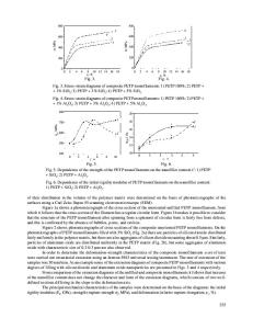
Carbon–carbon (C/C) composites are produced, among other processes, by chemical vapor infiltration (CVI): a heated fibrous preform is infiltrated by the chemical cracking of a vapor precursor of the matrix material inside the porous space of the perform.1 The quality of the materials manufactured by CVI relies on processing conditions (such as vapor precursor concentration, temperature and pressure), as well as on properties of the preform. Experimental determination of the conditions leading to optimal infiltration is long and expensive. That is why a global modeling of CVI is of great interest in optimizing the final porosity and homogeneity of the composites.2–6 This modeling requires sound knowledge of geometrical characteristics and transport properties of the perform.7–10 Numerous works have been performed to determine the internal surface area,11,12 thermal conductivity,13–18 binary gas diffusivity,9,19–22 Knudsen diffusivity,7,23–25 and the permeability26,27 of ideal media such as regular or random arrays of cylinders. However, real preforms exhibit a much more complex structure. Rather than modeling the porous medium by an arrangement of cylinders, this work aims at studying directly the preform by acquiring threedimensional (3D) images of it. X-ray tomography28 appears as an interesting technique for nondestructive characterization of 3D microstructure at different pore scales29–31
and has already been used successfully to characterize the structure of SiC fiber cloth lay-up preforms at macroporosity scale (pixel size of 15.6 m).32,33 However, applying this method to C/C composites at very high resolution is a challenging task, especially because the x-ray absorption contrast of high-resolution images of such light materials is faint, and the images are dominated by phase-contrast effects. In this study, tomography is used to image a C/C composite at two different scales (micro-and macro-porosity scale) and at three stages of infiltration. Geometrical characteristics (porosity, internal surface area, distribution of porosity, and pore sizes) are then determined from high-resolution images, and the results obtained are compared with experimental data and analytical results for model fiber structures. Also, an original method for determination of local fiber orientations in low-resolution images i
Data Loading...