Crystallization of ion beam deposited calcium phosphate coatings
- PDF / 1,565,798 Bytes
- 7 Pages / 576 x 792 pts Page_size
- 92 Downloads / 343 Views
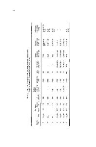
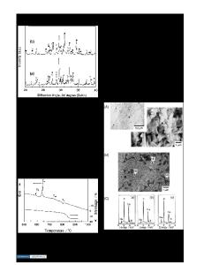
Amorphous calcium phosphate coatings on the order of 1 fim thick were deposited onto titanium and silicon substrates using an ion-beam sputtering technique. The target material utilized for sputter deposition was plasma-sprayed fluorapatite [Cai0(PO4)6F2; FA]. X-ray diffraction (XRD) and transmission electron microscopy (TEM) were employed to analyze the coatings. The amorphous as-deposited coatings were annealed in air (at 500 °C or 600 °C) to a crystalline state consisting of a polycrystalline FA matrix with a small amount of microcrystallites of a different composition. The higher annealing temperature (600 °C) tended to produce coarser FA and microcrystallite grains; however, the coatings buckled on the titanium substrates as a result of the heat treatment. Attempts to form the FA phase by in situ annealing in the vacuum chamber at a substrate temperature of 500 °C were not successful. The average bond strength for the as-deposited and 500 °C post-annealed coatings was comparable, while the lowest bond strength was observed in the 600 °C post-annealed coatings. The results suggest that the 500 °C post-annealed coatings have a suitable structure and possess sufficient adherence to be acceptable for use in certain medical and dental implant applications, and further tests under physiologic conditions will be conducted.
I. INTRODUCTION Calcium phosphate materials have drawn much attention over the years as coatings for biomedical implant devices. Hydroxylapatite [Ca10(PO4)6(OH)2; HA] and tricalcium phosphate [Ca 3 (PO 4 ) 2 ; TCP] are of particular interest because their chemical composition is similar to the mineral portions of bone. Metallic implants coated with a layer of calcium phosphate material, such as HA, have shown more significant and rapid interfacial attachment between the bone and implant than noncoated ones.1"4 The purpose of this study was to investigate another apatite material, fluorapatite [Ca10(PO4)6F2; FA], which has the potential for use in implant coating applications. A number of studies have shown that the presence of fluorine has certain beneficial effects in increasing the quantity and quality of bone formation in the body.5"9 Fluorapatite is structurally and chemically similar to HA, but is typically more stable during processing and in certain environments. This factor would make it potentially more useful for very thin sputter-deposited coatings which need good dissolution resistance and durability to function for a sufficient time in the human body. Although a great deal of effort has been concentrated on developing calcium phosphate coatings for implant materials, studies involving FA are still rare. Dhert et al. reported that titanium implants plasma sprayed with 50 fim thick FA had shown a high amount of bone apposition after 25 weeks of implantation in goats.10 1284
J. Mater. Res., Vol. 9, No. 5, May 1994
http://journals.cambridge.org
Downloaded: 13 Mar 2015
The plasma-sprayed FA coatings also gave a high pushout strength in bone, indicating possible application for certain
Data Loading...