Delayed and isolated abducens nerve palsy following minor head injury
- PDF / 526,081 Bytes
- 3 Pages / 595.276 x 790.866 pts Page_size
- 12 Downloads / 323 Views
LETTER TO THE EDITOR
Delayed and isolated abducens nerve palsy following minor head injury Jae Yeon Lee 1 & Yung Ju Yoo 1,2 Received: 24 September 2019 / Accepted: 18 January 2020 # Fondazione Società Italiana di Neurologia 2020
Introduction Unilateral abducens nerve palsy is reported to occur in 4.3% and bilateral injury in 2.1% of pediatric patients with head trauma [1]. Traumatic abducens nerve palsies occur immediately after trauma, and delayed onset of abducens nerve palsy after initial normal ocular deviation is rare. To the best of our knowledge, this is the first report on delayed and progressive bilateral abducens palsy following minor head trauma without intracranial lesion or skull fracture.
Case report An 11-year-old boy presented for evaluation with sudden onset of binocular horizontal diplopia, which appeared 2 days after a closed head injury; the patient had injured the posterior part of the head by falling on a concrete floor while playing outdoors and being pushed back by someone. The patient reported no loss of consciousness, and the initial neurologic examination was normal. Brain computed topography (CT) indicated no skull fracture or acute intracranial hemorrhage. He had no history of strabismus, eye patching, or ocular surgery. At presentation, the uncorrected visual acuities were 20/20 in both eyes (OU). Cycloplegic refraction revealed mild hyperopia OU. Funduscopic examination showed normal optic disc OU. There was no relative afferent pupillary defect, and color vision was normal OU. The alternate prism cover test (APCT) revealed 20 prism diopters (PD) of esotropia in the * Yung Ju Yoo [email protected] 1
Department of Ophthalmology, Kangwon National University School of Medicine, Chuncheon, South Korea
2
Department of Ophthalmology, Kangwon National University Hospital, #156, Baengnyeong-ro, Chuncheon 24289, South Korea
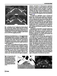
primary gaze at both distance and near fixation that increased to 30 PD in the right gaze. Ductions revealed − 1 deficit of abduction in the left eye but were otherwise full. The next day, the APCT showed 35 PD of esotropia, with a tendency to progress (Fig. 1). The limitation of abduction in the left eye was aggravated to − 3 degrees, and a − 1 deficit of abduction in the right eye was newly detected. For further evaluation, high-resolution thin section magnetic resonance imaging of the brain, including the abducens nerve, was performed and revealed no notable findings such as subarachnoid hemorrhage or gray or white matter abnormalities as also seen on the previous CT scan. The patient was treated symptomatically with occlusion of one eye to avoid binocular diplopia and was observed at 1-week intervals after being discharged. After 6 weeks of follow-up, the patient reported complete resolution of symptoms and his ductions and versions had returned to normal.
Discussion In the present case, bilateral abducens nerve palsy appeared 2 days after a minor head injury, and the angle of deviation increased progressively the next day. No other systemic or neurological symp
Data Loading...