Endovascular repair combined with staged drainage for the treatment of infectious aortic aneurysm: a case report
- PDF / 3,290,095 Bytes
- 4 Pages / 595.276 x 790.866 pts Page_size
- 53 Downloads / 316 Views
CASE REPORT
Open Access
Endovascular repair combined with staged drainage for the treatment of infectious aortic aneurysm: a case report Zhaoxiang Zeng1†, Zhenjiang Li1,2†, Yuxi Zhao1†, Junjun Liu1,3, Jiaxuan Feng1, Zaiping Jing1 and Rui Feng1*
Abstract Background: Infectious aortic aneurysm, defined as a focal dilation of an infectious arterial wall, is an uncommon life-threatening disease. Compared with open surgery, endovascular repair yields acceptable clinical outcomes. However, residual tissue infection may increase the risk of secondary intervention. Here, we present a successful case of endovascular repair combined with staged drainage for the treatment of infectious aortic aneurysm. Case presentation: A 58-year-old man presented to hospital with a 3-day history of lower back pain radiating to the back associated with fever. The dynamic imaging characteristics revealed rapid progress of infectious abdominal aortic aneurysm with negative blood culture. The patient underwent endovascular repair and salmonella enteritidis was identified through drain culture. Conclusions: Endovascular procedure and staged drainage can be feasible and effective option in selected cases. Keywords: Infectious abdominal aortic aneurysm, Aortitis, Pseudoaneurysm, EAVR, Case report
Background Infectious aortic aneurysm (or mycotic aortic aneurysm) is defined as a focal dilation of an infectious arterial wall. It is an uncommon life-threatening disease [1]. It is essential to establish an early diagnosis of infectious aneurysms for timely treatment to improve survival and long-term prognosis. Radiological imaging is critical for detecting initial aortic infection. However, the changes in early images may be minimal, making them easy to be neglected or mixed up [2]. We reported a case with complete sequential typical images of infectious abdominal aortic aneurysm in different stages recorded by series computed tomography (CT) scans, and the patient
* Correspondence: [email protected] † Zhaoxiang Zeng, Zhenjiang Li and Yuxi Zhao contributed equally to this work. 1 Department of Vascular Surgery, Changhai Hospital, Navy Medical University, Changhai Road 168#, Yangpu District, Shanghai, China Full list of author information is available at the end of the article
was successfully treated by endovascular procedure and staged drainage.
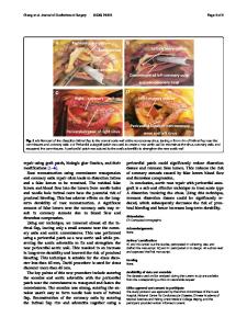
Case presentation A 58-year-old man presented to the emergency department with a 3-day history of lower back pain radiating to the back associated with fever (39.7 °C). The physical examination was unremarkable. He has a medical history of diabetes and current cigarette smoking. On admission, enhanced computed tomography angiography (CTA) revealed a moderate atherosclerotic infrarenal abdominal aorta with thickened aortic wall and periaortic fat stranding (Fig. 1a, 2a). A laboratory investigation showed that leukocyte count was normal, while the ration of lymphocytes decreased to 18.2% and eosinophil count was 0. The erythrocyte sedimentation rateand C-reactive peptide were elevated (34 mm/h and 138 mg/
Data Loading...