Evaluation of plantar fasciopathy shear wave elastography: a comparison between patients and healthy subjects
- PDF / 699,469 Bytes
- 6 Pages / 595.276 x 790.866 pts Page_size
- 1 Downloads / 415 Views
ORIGINAL PAPER
Evaluation of plantar fasciopathy shear wave elastography: a comparison between patients and healthy subjects Giuseppe Schillizzi1 · Federica Alviti2 · Chiara D’Ercole2 · Daniela Elia1 · Francesco Agostini2 · Massimiliano Mangone2 · Marco Paoloni2,3 · Andrea Bernetti2 · Patrizia Pacini1 · Giorgia Polti1 · Paolo Minafra4 · Vito Cantisani2 Received: 13 February 2020 / Accepted: 4 May 2020 © Società Italiana di Ultrasonologia in Medicina e Biologia (SIUMB) 2020
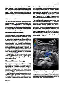
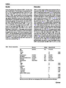
Abstract Purpose The aim of this study is to compare elasticity features between patients with plantar fasciitis (PFis) and an asymptomatic healthy control group using shear wave elastography (SWE) and to correlate SWE values with clinical scores. Methods Consecutive patients diagnosed with PFis and asymptomatic subjects were enrolled in the present study. Both groups underwent clinical, ultrasound (US), and SWE evaluation. A plantar fascia thickness > 4 mm was considered pathognomonic of PFis. SWE stiffness elasticity (Young’s modulus in kPa and shear wave velocity in m/s) was measured 1 cm distally from the calcaneal insertion. Correlations with VAS and the 17-Italian Foot Function Index (17-FFI) were determined. Results A total of 19 patients satisfied the inclusion criteria for the patient group and were enrolled in the study, and 21 healthy subjects were used as a control group. Statistically significant differences were found for shear wave velocity between the patient and the control group, with SWE findings of 3.8 (5.1; 1.5) m/s and 4.7 (4.07; 7.04) m/s, respectively (p = 0.006). Strong positive correlations were found between the SWE findings and both the pain and the functional scale (VAS: p = 0.001; FFI: p = 0.012). Conclusion SWE allows quantitative assessment of the stiffness of the plantar fascia and can show PFis alterations, increasing the diagnostic performance of B-mode US. In addition, SWE shows a strong correlation with clinical scores, improving patient assessment and follow-up. Keywords Shear wave elastography · Elastography · Elastosonography · Plantar fasciitis · Tendinopathy · Plantar fasciopathy · SWE
Introduction * Federica Alviti [email protected] Giuseppe Schillizzi [email protected] Vito Cantisani [email protected] 1
Deparment of Radiological, Oncological and AnatomyPathological Scinces “Sapienza” University, Rome, Italy
2
Department of Anatomy, Histology, Forensic Medicine and Orthopedics, Board of Physical Medicine and Rehabilitation, “Sapienza” University, Rome, Italy
3
Department of Physical Medicine and Rehabilitation, Azienda Policlinico Umberto I, Rome, Italy
4
Department of Radiology, Policlinico San Matteo, Pavia, Italy
Plantar fasciitis (PFis) is a common cause of inferior heel pain [1]. The typical symptom in patients with PFis is pain while taking the first steps in the morning or after a period of inactivity [2]. While standing or during other daily routine activities, patients often experience a progressive worsening of symptoms, with increased c
Data Loading...