F-18 FDG PET/CT in Bilateral Diffuse Pulmonary Lymphangitic Carcinomatosis
- PDF / 137,012 Bytes
- 2 Pages / 595.276 x 790.866 pts Page_size
- 24 Downloads / 307 Views
INTERESTING IMAGE
F-18 FDG PET/CT in Bilateral Diffuse Pulmonary Lymphangitic Carcinomatosis Raja Senthil & Rahul Parghane & Raghava Kashyap & Anish Bhattacharya & Bhagwant Rai Mittal
Received: 1 September 2011 / Revised: 12 December 2011 / Accepted: 16 January 2012 / Published online: 4 February 2012 # Korean Society of Nuclear Medicine 2012
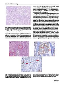
A 51-year-old female patient, who had undergone left-sided modified radical mastectomy for left breast carcinoma 4 years ago, presented with dyspnea of 4 months duration. F-18 FDG PET/CT of this patient showed diffusely increased FDG uptake in the bilateral lung fields along the thickened bronchovascular bundles (Fig. 1a–c). SUVmax of lymphangitic lung was 5.2. The standardized uptake ratio (SUR) of mediastinal blood pool to lymphangitic lung was 0.44. High resolution computed tomography (HRCT) of the same patient showed thickening of interlobular septa and bronchovascular bundles, with preservation of normal parenchymal architecture. Multiple intrapulmonary nodules and bilateral hilar lymphadenopathy were also seen (Fig. 1d, e). These findings are consistent with pulmonary lymphangitic carcinomatosis (PLC). The lungs are the second most common sites for metastases after lymph nodes [1]. These metastases are usually
R. Senthil : R. Parghane : R. Kashyap : A. Bhattacharya : B. R. Mittal (*) Department of Nuclear Medicine & PET, Postgraduate Institute of Medical Education and Research, Chandigarh, India e-mail: [email protected]
nodular on radiologic images. PLC with interstitial involvement constitutes only 7% of pulmonary metastases [2]. The most common primary sites, in order of frequency, are adenocarcinoma of the lung, breast, stomach, colon, and prostrate [2]. HRCT has been the modality of choice in the radiologic diagnosis of PLC. Only a few studies have described the F-18 FDG PET/CT findings in pulmonary lymphangitic carcinomatosis [3–5]. These studies have shown diffusely increased FDG uptake corresponding to the typical changes in the CT as the most common finding. One study has reported that F-18 FDG PET/CT is 100% specific and 86% sensitive in diagnosing PLC by subjective analysis. The mean SUV in the region of pulmonary lymphangitic carcinomatosis was 1.37±0.64 vs. 0.51±0.29 in normal lung. The SUR of mediastinal blood pool to lymphangitic lung was 1.26±0.45, and that of blood pool to normal lung was 3.78±1.37 [5].
Nucl Med Mol Imaging (2012) 46:150–151
151
Fig. 1 F-18 FDG PET/CT images (A, B, C) showing diffusely increased FDG uptake in the bilateral lung fields along the thickened bronchovascular bundles. High resolution computed tomography (D, E) of the same patient
showed thickening of interlobular septa and bronchovascular bundles, with preservation of normal parenchymal architecture. Multiple intrapulmonary nodules and bilateral hilar lymphadenopathy are also seen
Conflict of interest We declare that we have no conflict of interest.
3. Digumarthy SR, Fischman AJ, Kwek BH, Aquino SL. Fluorodeoxyglucose positron emission tomography pattern of pulmonar
Data Loading...