Nanometer-Scale Modification and Imaging of Polyimide Films by Scanning Force Microscopy
- PDF / 2,232,456 Bytes
- 6 Pages / 420.48 x 639 pts Page_size
- 69 Downloads / 330 Views
NANOMETER-SCALE MODIFICATION AND IMAGING OF POLYIMIDE FILMS BY SCANNING FORCE MICROSCOPY
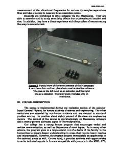
W. N. UNERTL and X. JIN University of Maine, Orono, Maine 04469 ABSTRACT The sharp tip of a scanning force microscope can be used to make controlled modifications of polymer surfaces. In this paper, we describe the properties of micrometer size pits up to 900 A deep formed on Kapton-H surfaces. The structure at the bottom of the pits appears to be closely related to the degree of crystallinity near the surface. We also use elasticity theory to estimate that the resolution of scanning force microscopy for polymer surfaces is about 160 A for tips with 400 radius. This estimate agrees well with the resolution obtained in images of polyirnide surfaces. INTRODUCTION The scanning force microscope (SFM) is a new technique to study the surface structure and surface mechanical properties of nonconducting materials with nanometer scale resolution [1,2]. This paper describes a study using the SFM tip to create micropits on the polyimide, Kapton-H [3]. Kapton-H is widely used in electronic packaging as a thin film dielectric material [3,41; it is made from the monomers pyromellitic dianhydride (PMDA) and oxydianylene (ODA). In packaging, a number of material's properties including surface and near surface homogeneity, adhesion, and wear are important and can be studied with the SFM. Previously, we have described the properties of sub-micrometer wide grooves that were scribed in a polyimide surface with the SFM tip [5]. First, we briefly describe the force microscope and discuss its resolution for the case of soft adhesive materials. Next, we present examples of micrometer size pits formed on polyimide surfaces by raster scanning the SFM tip under an applied load. Finally, we discuss the need for a more detailed understanding of the surface mechanical properties before the controlled modifications can be exploited as a means of extracting information about the surface properties of polyimides and other soft materials. THE SURFACE FORCE MICROSCOPE AND SAMPLES The commercial SFM [6] used for this study is shown schematically in Fig. 1. The sample is mounted on top of a cylindrical piezoelectric tube scanner (PZT scanner) that controls the z-displacement between the sample and tip and also xy-raster scans the sample over a desired area for imaging. The probe tip is a Si 3 N 4 pyramid 2.6 pm high with a 4 pm base and a tip radius R specified by the supplier [6] to be less than 40 nm. This tip is an integral part of a micro-fabricated Si 3 N 4 cantilever; the cantilevers used for the measurements described below had a force constant k of 0.37 N/m. The sample is brought into contact with the S13 N4 tip at a predetermined applied load W. W is determined from the tip displacement Az and k using Hooke's law, W = -kAz. The tip displacement is measured using the beam deflection method shown in Fig. 1. Light from a laser diode is focused on the cantilever tip and the reflected light collected by a position sensitive Mat. Res. Soc. Symp. Proc. Vol. 239
Data Loading...