Radiotherapy for recurrent central Giant cell granuloma: a case report
- PDF / 856,017 Bytes
- 4 Pages / 595.276 x 790.866 pts Page_size
- 87 Downloads / 332 Views
CASE REPORT
Open Access
Radiotherapy for recurrent central Giant cell granuloma: a case report Qi Zhang1,2†, Zelai He1,2†, Gengming Wang1,2 and Hao Jiang1,2*
Abstract Background: Central giant cell granuloma (CGCG) is a rare, non-neoplastic, benign lesion that exhibits expansive and osteolytic biological behavior. CGCG treatment and management is challenging for clinicians. Case presentation: This report presents the treatment and management of recurrent, aggressive CGCG after surgical resection. After informed consent was obtained, the patient underwent radiotherapy. The lesion size was reduced significantly, with no evidence of recurrence or malignant transformation. Conclusions: This treatment experience indicates that radiotherapy can be used as a rescue treatment for complicated CGCG involving vital neurovascular structures of the cranial base. Keywords: Central giant cell granuloma, Radiotherapy, Surgical resection
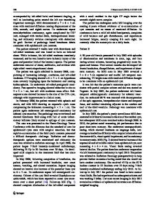
Background Central giant cell granuloma (CGCG) is a rare, non-neoplastic, benign lesion that exhibits expansive and osteolytic biological behavior [1]. The lesion principally affects the mandible and maxilla and rarely affects the skull or small bones of the hands and feet [2, 3]. To the best of our knowledge, the present case is the first report of recurrent CGCG of the nasal cavity and sinuses and its successful treatment by intensity-modulated radiotherapy. Clinical presentation The patient, a 35-year-old man without a previous history of trauma or predisposing factors, was initially referred to the Otorhinolaryngology Department in September 2014 for a neoplasm in the right nasal cavity. Surgical resection of the neoplasm was performed. The postoperative pathological findings supported the diagnosis of CGCG (Fig. 1d). After surgical resection, clinical and radiographic examinations revealed complete remission. The patient was referred to our center in December 2015 for massive facial swelling (Fig. 1e). The patient’s main complaint was “an * Correspondence: [email protected] † Qi Zhang and Zelai He contributed equally to this work. 1 Department of Radiation Oncology, The First Affiliated Hospital of Bengbu Medical University and Tumor Hospital Affiliated to Bengbu Medical University, 287 Changhuai Road, Bengbu, Anhui 233004, People’s Republic of China 2 Anhui Province Key Laboratory of Translational Cancer Research Affiliated to Bengbu Medical University, Bengbu 233004, China
intermittent headache lasting for more than three months, accompanied with a progressive decline in hearing lasting for one month”. Computed tomography (CT) revealed that the lesion affected the right nasal cavity, maxillary sinus, right frontal lobe, right eye, and skull base bone (Fig. 1b). Magnetic resonance imaging (MRI) showed a large lesion reaching 9.8 × 7.6 cm in size. The lesion primarily involved the nasal cavity, nasopharynx, and bilateral ethmoid sinus. Additionally, the invasive lesion expanded intracranially, affecting the eyes (bilateral), extraocular muscles, and frontal lobe of the parenchyma. Addi
Data Loading...