Speculation on the reconstruction of vicinal {100} faces of vapor-deposited diamond
- PDF / 319,324 Bytes
- 3 Pages / 576 x 792 pts Page_size
- 43 Downloads / 261 Views
Recent morphological studies of vapor-deposited diamond indicate (1) that growth on the diamond {100} surface is mediated by steps and (2) that some defects cause rapid initiation of new layers, resulting in characteristic pyramids with edges parallel to surface (110) directions. If growth on the diamond {100} surface does indeed occur at steps, knowledge of their atomic structure is essential to an understanding of diamond growth. We reexamine results of a recent STM study of homoepitaxial diamond films and suggest that the surfaces on which growth occurs consist of regularly spaced double height steps separated by integral numbers of dimer rows running parallel to the step edges.
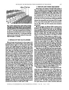
Both low energy electron diffraction (LEED) and direct imaging scanning tunneling microscopy (STM) and atomic force microscopy (AFM) reveal a 2 X 1 reconstruction of the {100} face of vapor-deposited diamond.1"3 Correspondingly, some models of the chemical vapor deposition (CVD) process assume growth on this dimerized but otherwise flat surface.4'5 Morphological studies using both electron microscopy and scanning tip microscopies (STM and AFM) strongly suggest that growth on the diamond {100} surface actually occurs at steps.6"8 A realistic growth model must begin with an accurate picture of the surface on which growth actually occurs. Unfortunately, diffraction techniques that average over large areas cannot readily provide such information from the complex, highly defective diamond surfaces currently available. Direct atomic resolution imaging has been achieved only on (almost) atomically flat regions of low index diamond surfaces. In this note, we reexamine results of a recent STM study of the morphology of diamond homoepitaxy to infer the atomic structure of the growth steps. Ravi and Joshi first reported the appearance of "ledges" on the diamond {100} surface and interpreted the observation in terms of growth by lateral epitaxy at steps or ledges.6 Okada and co-workers also report steplike features, pyramidal pits, and what appear to be growth spirals, all of which they interpret in terms of step-mediated growth.7 Recent higher-resolution STM and AFM studies reveal similar structures, as well as ubiquitous pyramidal features which will be the focus of this note.8'9 As illustrated by Fig. 1, the pyramids are quite shallow with abrupt and distinct changes in slope. (A more descriptive term might be "ziggurat.") They are always aligned with their edges parallel to (110) directions in the {100} face. Although some pyramids have a sharp peak, most exhibit a central triangular feature. These have been identified by Sutcu et al. as 1770 http://journals.cambridge.org
J. Mater. Res., Vol. 8, No. 8, Aug 1993
Downloaded: 14 Mar 2015
FIG. 1. Scanning electron microscope image of a typical pyramidal structure on a {100} oriented homopepit-axial diamond film. This pyramid is approximately 6 ixm wide across the base with its edges parallel to surface (110} directions.
penetration twins9 but were simply termed "gross defects" by Everson and Ta
Data Loading...