Three-Dimensional PEG Hydrogel Construct Fabrication using Stereolithography
- PDF / 256,722 Bytes
- 7 Pages / 612 x 792 pts (letter) Page_size
- 85 Downloads / 278 Views
L5.5.1
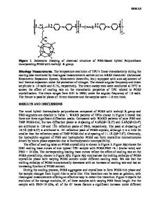
Three-Dimensional PEG Hydrogel Construct Fabrication using Stereolithography Karina Arcaute1, Luis Ochoa1, Frank Medina1, Chris Elkins2, Brenda Mann3 and Ryan Wicker1 1 W.M. Keck Border Biomedical Manufacturing and Engineering Laboratory, Mechanical Engineering, University of Texas at El Paso, El Paso, TX, USA. 2 Mechanical Engineering and Radiology, Stanford University, Stanford, CA, USA. 3 Sentrx Surgical, Inc., Salt Lake City, UT, USA. ABSTRACT Layered manufacturing (LM) using stereolithography (SL) of aqueous polymer solutions was accomplished so three-dimensional (3D) tissue engineered scaffolds with complex distributions of bioactive agents could be produced. Successful LM with embedded channel architectures required investigation of hydrogel thickness or cure depth as a function of photoinitiator type and concentration, energy dosage, and polymer concentration in solution. Poly(ethylene glycol) dimethacrylate (PEG-dma) with an average molecular weight of 1000 in quantities of 20% and 30% (w/v) was prepared in distilled water. Different concentrations of two photoinitiators (PIs), Sarcure1121 (2-hydroxy-2-methyl-1-phenyl-1-propanone) and Irgacure 2959 (2-hydroxy-1-[4-(2-hydroxyethoxy)phenyl]-2-methyl-1-propanone), were used to vary gel thickness at select energy dosages by controlling the scan speed of the SL machine’s ultraviolet scanning system. Gel thickness was a strong function of PI type and concentration, energy dosage, and PEG-dma concentration, especially at the low PI concentrations required for implantation. The gel thickness curves were utilized to demonstrate LM for two construct geometries where different layer thicknesses were required to successfully fabricate the constructs. This work demonstrates the effective use of SL as a processing technique for complex 3D tissue scaffolds and addresses some practical considerations associated with the use of hydrogels in LM. INTRODUCTION One of the approaches used in tissue engineering (TE) relies on the use of a scaffold to provide the mechanical support and structure for cells to grow and regenerate a damaged tissue. Researchers have investigated a wide variety of strategies for engineering implantable scaffolds for tissue regeneration. The strategies depend on the material used to fabricate the scaffold and its intended application [1, 2]. Layered manufacturing (LM) or rapid prototyping (RP) technologies have been employed quite extensively for manufacturing tissue scaffolds. Although RP was first commercialized in the mid 1980s, the use of RP in TE was somewhat recent, especially in soft tissue applications, with activity increasing at a rapid rate. RP is generally becoming accepted as the most capable manufacturing method for TE, since by its LM nature, control over scaffold characteristics as well as placement of cells and bioactive agents within the scaffold are possible. Stereolithography (SL) is one of the RP processes used in TE [3, 4, 5]. Commercial SL works by drawing two-dimensional (2D) patterns with a focused ultraviolet (UV) laser
Data Loading...