Ultrasound diagnosis of bilateral ectopic pregnancy: Still a myth?
- PDF / 873,654 Bytes
- 3 Pages / 595.276 x 793.701 pts Page_size
- 0 Downloads / 377 Views
Hellenic Journal of Surgery (2015) 87:6, 490-492
Ultrasound Diagnosis of Bilateral Ectopic Pregnancy: Still A Myth? P. Sinha, A. Koumousidis, M. Carr, C. Sofoudis
Abstract Bilateral ectopic pregnancy is a rare entity. If missed, it can lead to significant increase in morbidity of the patient. Our study highlights the importance of imaging in aiding in the diagnosis of this type of extrauterine pregnancy as well as the necessity of a prompt evaluation of both adnexae during laparoscopy. Key words: Ectopic pregnancy, fallopian tubes, bilateral, transvaginal ultrasound scan, laparoscopy
Introduction Ectopic pregnancy occurs in 0.5-2% of all pregnancies and a double ectopic pregnancy is very rare [1]. Spontaneous bilateral ectopic pregnancy is difficult to diagnose preoperatively, indicating the limitations of ultrasonography. Laparoscopy has always been considered as gold standard in diagnosing this rare condition. However, early diagnosis is essential to avoid potential fatal complications and preserve future fertility.
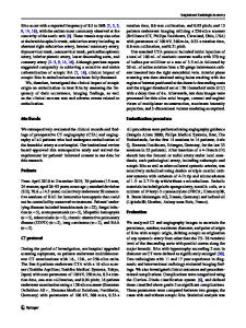
Case Presentation We report the case of a 30-year-old woman who presented to the accident and emergency department (A & E) with severe abdominal pain and persistent diarrhoea. Her serum beta chorionic gonadotropin (β-hCG) was more than 70,000 mIU/mL and progesterone was 70 ng/ml. An ultrasound scan confirmed live ectopic pregnancy in the left adnexa (Figure 1) and a large haematosalpinx on the right side with a possible ectopic pregnancy (Figure 2). Due to the urgency of the case, the patient was prepared for theatre: the necessary bloods were taken (full blood count, group and save), the patient was debriefed and her signed informed consent was obtained. Four units of blood were cross-matched. The patient was transferred to the operating theatre for surgical management of a partially ruptured
ectopic pregnancy (laparoscopy, +/- exploratory laparotomy, +/- salpingostomy, +/- salpingectomy, +/- oophorectomy). The laparoscopic findings were highly suggestive of bilateral ectopic pregnancy; consequently, bilateral salpingectomy was performed. The histopathology examination confirmed the presence of two gestational sacs. One sac was within the tube (Figure 3, 4) and another extruded out of the tube among the blood clots extracted from the POD. Within one of the gestation sacs was what appeared to be an anencephalic foetus of 15 mm in length.
Discussion The incidence of bilateral tubal ectopic pregnancies is reported to range from 1/725 to 1/1,580 extrauterine pregnancies [2]. As its incidence is rising due to the increase in
P. Sinha, A. Koumousidis, M. Carr OBS/GYN C.S ofoudis Scientific associate, OBS/GYN East Sussex Hospital NHS Trust, St Leonards-on-Sea, England Corresponding author: Dr. Antonios Koumousidis 43- 46 Oast House Close, St. Leonards on Sea, East Sussex, England, TN377UY Tel.: +44 07449829965, e-mail: [email protected] Received 29 Aug 2015; Accepted 10 Oct 2015
Hellenic Journal of Surgery 87
Figure 1. Transvaginal ultrasound showing the gestational sac and the foetal pole at
Data Loading...