Variation of Stratum Corneum Biophysical and Molecular Properties with Anatomic Site
- PDF / 312,053 Bytes
- 7 Pages / 595.276 x 790.866 pts Page_size
- 43 Downloads / 355 Views
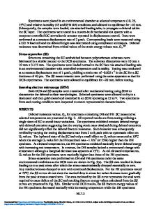
Research Article Variation of Stratum Corneum Biophysical and Molecular Properties with Anatomic Site Diar Mohammed,1 Paul J. Matts,2 Jonathan Hadgraft,1 and Majella E. Lane1,3
Received 30 June 2012; accepted 2 August 2012; published online 18 August 2012 Abstract. Several serine protease enzymes are known to be involved in both normal desquamation and the inflammatory processes of the skin. Alteration in the activity of these proteases should also affect corneocyte maturity and size as well as stratum corneum thickness. The aim of the present work was to characterise the baseline changes in corneocyte size, corneocyte maturity, selected protease activity (specifically, Kallikreins-5 and 7, tryptase), protein content and trans-epidermal water loss (TEWL) as a function of anatomic site. The anatomic sites investigated were: cheek, abdomen, wrist and mid-ventral forearm. TEWL values were highest for the cheek (p0.1).
Fig. 2. TEWL measurements for different anatomic sites (mean± SEM, n=22)
SC Protein Measurement Figure 3 shows the mean protein content per strip (for tape strips 2–4) data for different anatomic sites. Apart from wrist versus abdomen (p0.05). The data and SEM values are in line with previous values we have reported (11). There were no significant differences in the amount of protein removed for males compared with females (p>0.1) when categorising the volunteers according to gender (data not shown). When the data were analysed by ethnic group (data not shown), similar trends were observed for both Caucasians and Black subjects, i.e. no significant differences in protein content removed from various anatomical sites were noted (p>0.1).
Corneocyte Maturity and Surface Area The results for corneocyte maturity, for tape strip 1, when analysed as a function of anatomic site, are shown in Fig. 4. There are no differences in overall corneocyte maturity between males and females or between ethnic groups (data not shown, p>0.1). The corneocyte maturity of the cheek is the lowest followed by wrist then mid-ventral forearm and abdomen; however, cheek and wrist were not significantly different (p> 0.05). Values for cheek and wrist are significantly (p0.1) from the abdomen. For the two ethnic groups, a similar trend was observed, i.e. reduced corneocyte size for the cheek compared with the mid-ventral forearm or abdomen, but there were no significant differences in corneocyte surface area between the groups (p>0.05).
Fig. 3. Mean SC protein content removed per strip from different anatomic sites, tape strips 2–4 (n=22, mean±SEM)
Variation of Stratum Corneum Biophysical and Molecular Properties
809
Fig. 4. Corneocyte maturity at different anatomic sites, tape strip 1 (n=22, mean±SEM)
Fig. 6. Correlation between corneocyte maturity and surface area (n=22, mean±SEM)
A positive correlation between corneocyte maturity and surface area over the sites studied is also evident (Fig. 6), i.e. the greater the maturity at the specific site, the larger the corneocyte size. A similar correlation between corneocyte maturity and
Data Loading...