Surface Modification of Superparamagnetic Nanoparticles for in-vivo Bio-medical Applications
- PDF / 515,905 Bytes
- 6 Pages / 612 x 792 pts (letter) Page_size
- 72 Downloads / 284 Views
Surface modification of superparamagnetic nanoparticles for in-vivo bio-medical applications D. K. Kim1, M. Toprak1, M. Mikhailova1, Y. Zhang1, B. Bjelke2, J. Kehr3, M. Muhammed1. 1
Materials Chemistry Division, Royal Institute of Technology, SE-100 44 Stockholm, Sweden MRI-Center, Experimental Unit, Karolinska Institutet, SE-171 76 Stockholm, Sweden 3 Division of Cellular and Molecular Neurochemistry, Karolinska Institutet, SE-171 77 Stockholm, Sweden 2
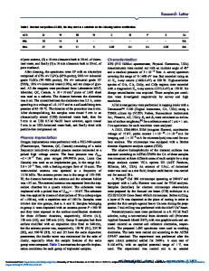
ABSTRACT Chemical modifications of Superparamagnetic Iron Oxide Nanoparticles (SPION) surfaces by attachment of functional groups and further covalent coupling with biodegradable substances have been studied. Based on computer-assisted chemical equilibrium calculations, several optimum operation conditions for a coprecipitation process of magnetite nanoparticles were predicted. These particles were immobilized by ultra-thin films of PVA, Dextran, Dextrin, PEG and MPEG to obtain a biocompatible particle surface for further functionalization purposes. The effect of surface modification of the superparamagnetic nanoparticles in terms of chemical and physical properties of the samples was investigated with several techniques, including microelectrophoresis measurement. The feasibility of using SPION in biomedical applications was investigated by in-vivo treatment in rat brains. INTRODUCTION Application of SPION for hyperthermia of biological tissue has been known in principle for more than four decades [1]. Especially, for MR imaging purposes, imaging agents are required to increase conspicuity of adjacent internal organs and tissue. Moreover, SPION can be used to monitor extracellular macromolecules both at a single cell level (genes and proteins) and at a network level (intercellular communication) by in-vivo monitoring of particle movement in the living brain tissue. Biomedical applications using SPION require narrow size distribution and surface modifications with biocompatible materials, i.e. nonimmunogenic, nonantigenic, and protein-resistant. To transport this hydrophilic substance in biological membrane system, surface modification of the nanoparticle was necessary to adjust the zeta potential close to zero. The influence of physico-chemical characteristics on the uptake of particles by the mononuclear phagocyte system (MPS) and accumulation in the reticuloendothelial system, comprising mainly of the macrophages of the liver and the spleen has been reported. [2] Also, ferrofluids composed of SPION are not only affected by an inhomogeneous particle size distribution, but also by the surface charge of the particles in the solution. One of the practical methods to stabilize the ferrofluids is through electrostatic stabilization, achieved by the repulsion of equally charged surface. The repulsive force results from creation of an electric double layer around the particles, which is dependent on dispersion of the particles into a polar media, pH, concentration, and ionic strength of the suspension. Specific medical application of these particles also requires a mod
Data Loading...