Visual Field Changes in Glaucoma
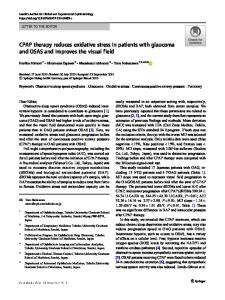
Figure 34.1 shows the different defects that can be found in the visual field of a glaucoma patient.
- PDF / 2,249,006 Bytes
- 17 Pages / 504.57 x 720 pts Page_size
- 55 Downloads / 302 Views
Contents
34.1
34.1
Paracentral and Pericentral Scotomas..... 513
34.2
Scotomas in the Circular Area Between 10° and 20° (Isolated from the Blind Spot) .................................. 515
34.3
Exclusion of the Blind Spot ....................... 515
34.4
Arcuate Scotoma ........................................ 515
34.5
Ronne’s Step ............................................... 517
34.6
Practical Classification of the Visual Fields Obtained in the Goldmann Perimeter .................................................... 518
34.7 34.7.1 34.7.2 34.7.3
Normal Field............................................... Grade I ......................................................... Grade II ........................................................ Grade III.......................................................
34.8
Correlation Between Visual Field Alterations and the Optic Disk ................. 520
34.9
Optic Disk Drusen or Hyaline Verrucosities ............................................... 523
34.10
Papillary Pit ................................................ 526
34.11
Summary..................................................... 526
518 518 519 519
References ................................................................. 529
34
Paracentral and Pericentral Scotomas
Figure 34.1 shows the different defects that can be found in the visual field of a glaucoma patient. Visual field alterations begin with a series of small isolated scotomas in the circular area between 10° and 20° surrounding the central point, above and below the physiological scotoma (blind spot) without being related to it (Fig. 34.1). In my experience, exclusion of the blind spot below 50 years of age, with 10/10 vision, is also an early manifestation of the disease. Later, these small scotomas join together to form a larger scotomas: the arcuate or Bjerrum’s scotoma, which can come to have the shape of a comet, with its thinnest point close to the blind spot. It stops abruptly on the nasal side because of the arrangement of the retinal fibers. Usually, the upper one is the first to appear. A lower arcuate scotoma forms in the same way. When both are well developed, they meet in the nasal field and, from the arrangement of the retinal fibers in the raphe, form a step called Ronne’s step. Thus, a central island of vision remains, limited by a scotoma called a ring scotoma, with a central edge which is seen to be abrupt in static perimetry and an outer edge that is generally a gentle slope. At the same time, a loss of nasal field can occur, to a greater or lesser extent, the
R. Sampaolesi et al., The Glaucomas, DOI 10.1007/978-3-642-35500-4_34, © Springer-Verlag Berlin Heidelberg 2014
513
34
514
a
b
c
d
Visual Field Changes in Glaucoma
e
Fig. 34.1 Typical visual field lesions in glaucoma. (a) 1 Small isolated scotomas in the circular area between 10° and 20°. (b) 2 Bjerrum’s arcuate scotoma originated by the union of earlier ones. (c) 3 Inferior Bjerrum’s arcuate scotoma. (d)
4 Union of inferior arcuate scotoma w
Data Loading...