Visualization of Enthesitis by Ultrasound: a Key Diagnostic Tool in Spondyloarthropathy Diagnosis and Management
- PDF / 1,717,113 Bytes
- 6 Pages / 595.276 x 790.866 pts Page_size
- 21 Downloads / 292 Views
IMAGING (J SAMUELS, SECTION EDITOR)
Visualization of Enthesitis by Ultrasound: a Key Diagnostic Tool in Spondyloarthropathy Diagnosis and Management Gurjit S. Kaeley 1
# Springer Science+Business Media, LLC, part of Springer Nature 2020
Abstract Purpose of Review The purpose of this review is to report on the current status of evaluation of enthesitis with sonography. Sonography can visualize peripheral entheses in great detail since most of them are superficial. Visualization of entheses has provided insights into the pathophysiology of enthesitis and highlighted the interconnectivity of stress dissipation mechanisms about the enthesis. The primary use of entheseal sonography presently revolves around diagnostic use. Recent Findings There is increasing awareness of biomechanical confounders that lead to entheseal changes, which are more prevalent in males, patients with higher body mass index, and those who are more active—though there are several approaches to mitigate biomechanical confounders in sonographic evaluation. Summary Entheseal sonography may also provide prognostic information in predicting the severity of axial syndesmophyte formation. Although entheseal outcome scoring systems are available, there is sparse data on its use as an outcome tool, and hence, this is a fertile area for future research. Keywords Enthesitis . Sonography . Ultrasound . Spondyloarthropathy . Biomechanical confounders . Fibromyalgia
Introduction Enthesitis is a key pathophysiologic finding in the spondyloarthropathies, such as psoriatic arthritis (PsA), ankylosing spondylitis (AS), reactive arthritis, and inflammatory bowel disease-associated arthritis. Recent articles review the histological concept of non-axial entheses as predominantly fibrocartilaginous entheses [1, 2]. In summary, entheses function not only as anchor points to promote movement but have necessary shock-absorbing capacities. Anatomic structures that allow force dissipation include the spring-like formation of collagen in tendons, the fibrocartilaginous transition zone from tendon to bone, and adjacent fat pads and bursae. Recognition that these structures rather than just the mere tendon or entheses attachment to the bone may be involved in enthesitis has given rise to the enthesis organ concept This article is part of the Topical Collection on Imaging * Gurjit S. Kaeley [email protected] 1
Division of Rheumatology, University of Florida College of Medicine, 653-1 West Eight Street, LRC 2nd Floor L-14, Jacksonville, FL 32209-6561, USA
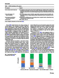
(Fig. 1). Sonography utilizes non-ionizing sound waves to image peripheral entheses. Since the majority of these are superficial, high fidelity imaging allows for a detailed evaluation of multiple components of the entheses structure (Fig. 2, Table 2). The section below will highlight essential differences in the selection of anatomic structures used in sonographic entheseal indices, biomechanical confounding factors, and critical limitations in the diagnostic use of these instruments.
Diagnostic Use of Enthesea
Data Loading...