Nonlinear optical microscopy is a novel tool for the analysis of cutaneous alterations in pseudoxanthoma elasticum
- PDF / 952,670 Bytes
- 10 Pages / 595.276 x 790.866 pts Page_size
- 38 Downloads / 314 Views
ORIGINAL ARTICLE
Nonlinear optical microscopy is a novel tool for the analysis of cutaneous alterations in pseudoxanthoma elasticum Norbert Kiss 1,2 & Luca Fésűs 1,2 & Szabolcs Bozsányi 1,2 & Flóra Szeri 3 & Matthias Van Gils 4,5 & Viktória Szabó 6 & Anikó Ilona Nagy 7 & Bernadett Hidvégi 1 & Róbert Szipőcs 2,8 & Ludovic Martin 9 Olivier Vanakker 4,5 & Tamás Arányi 10 & Béla Merkely 7 & Norbert M. Wikonkál 1 & Márta Medvecz 1
&
Received: 18 February 2020 / Accepted: 16 April 2020 # The Author(s) 2020
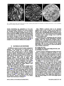
Abstract Pseudoxanthoma elasticum (PXE, OMIM 264800) is a rare autosomal recessive disorder with ectopic mineralization and fragmentation of elastin fibers. It is caused by mutations of the ABCC6 gene that leads to decreased serum levels of inorganic pyrophosphate (PPi) anti-mineralization factor. The occurrence of severe complications among PXE patients highlights the importance of early diagnosis so that prompt multidisciplinary care can be provided to patients. We aimed to examine dermal connective tissue with nonlinear optical (NLO) techniques, as collagen emits second-harmonic generation (SHG) signal, while elastin can be excited by two-photon excitation fluorescence (TPF). We performed molecular genetic analysis, ophthalmological and cardiovascular assessment, plasma PPi measurement, conventional histopathological examination, and ex vivo SHG and TPF imaging in five patients with PXE and five age- and gender-matched healthy controls. Pathological mutations including one new variant were found in the ABCC6 gene in all PXE patients and their plasma PPi level was significantly lower compared with controls. Degradation and mineralization of elastin fibers and extensive calcium deposition in the mid-dermis was visualized and quantified together with the alterations of the collagen structure in PXE. Our data suggests that NLO provides high-resolution imaging of the specific histopathological features of PXE-affected skin. In vivo NLO may be a promising tool in the assessment of PXE, promoting early diagnosis and follow-up. Keywords Pseudoxanthoma elasticum . Nonlinear optical microscopy . Multiphoton microscopy . Elastin . Calcification
Introduction Pseudoxanthoma elasticum (PXE, OMIM#264800) is a rare autosomal recessive connective tissue disorder characterized by
ectopic mineralization and fragmentation of elastin fibers. At the genetic level, homozygous or compound heterozygous lossof-function mutations in the ABCC6 gene (OMIM#603234) are the main cause of PXE [1]. ABCC6 encodes an ATP binding
Norbert M. Wikonkál and Márta Medvecz share senior authorship. * Márta Medvecz [email protected]
5
Department of Biomolecular Medicine, Ghent University, Ghent, Belgium
6
Department of Ophthalmology, Semmelweis University, Budapest, Hungary
7
Heart and Vascular Center, Semmelweis University, Budapest, Hungary
1
Department of Dermatology, Venereology and Dermatooncology, Semmelweis University, 41 Mária Street, Budapest H-1085, Hungary
2
Wigner RCP, Institute for Solid State Physics and Optics
Data Loading...