Primary Small Cell Neuroendocrine Carcinoma of the Kidney: Morphological, Immunohistochemical, Ultrastructural, and Cyto
- PDF / 755,320 Bytes
- 11 Pages / 595.276 x 790.866 pts Page_size
- 107 Downloads / 284 Views
Primary Small Cell Neuroendocrine Carcinoma of the Kidney: Morphological, Immunohistochemical, Ultrastructural, and Cytogenetic Study of a Case and Review of the Literature Stefano La Rosa & Barbara Bernasconi & Donata Micello & Giovanna Finzi & Carlo Capella
Published online: 19 December 2008 # Humana Press Inc. 2008
Abstract Poorly differentiated neuroendocrine carcinomas (PDNECs) of the kidney are extremely rare high-grade cancers accounting for only 42 cases reported in the literature. In this paper, we describe the morphological, immunohistochemical, ultrastructural, and for the first time, cytogenetic features of a renal PDNEC. In addition, we have reviewed the literature and compared the published clinicopathological data with our morphological and genetic results. The tumor arose within the kidney parenchyma and showed the typical histological features of a pure small cell PDNEC. Fluorescence in situ hybridization study demonstrated a complex chromosomal assessment indicative of a high degree of chromosome instability with gain of multiple chromosomes, loss of p53, and amplification of myc gene. These results suggest that renal PDNEC has a different genetic background to renal clear cell carcinoma, mainly characterized by the loss of the short arm of chromosome 3. Conversely, genetic alterations seem to resemble those of type 2 papillary renal cell carcinoma. The review of the literature demonstrated that PDNECs are associated with poor prognosis and that parenchymal tumors show some differences from those S. La Rosa (*) : G. Finzi Department of Pathology, Ospedale di Circolo, Viale Borri 57, 21100 Varese, Italy e-mail: [email protected] B. Bernasconi : D. Micello : C. Capella Department of Human Morphology, Anatomic Pathology Unit, University of Insubria, Varese, Italy C. Capella Centro Insubre di Biotecnologie per la Salute Umana, Varese, Italy
arising in the pelvis, in that parenchymal tumors are purely neuroendocrine while pelvic tumors are mostly mixed neuroendocrine–exocrine neoplasms. Keywords poorly differentiated neuroendocrine carcinoma . small cell carcinoma . kidney . FISH . cytogenetic
Introduction Poorly differentiated neuroendocrine carcinomas (PDNECs) of small, intermediate, and large cell types are high-grade epithelial tumors associated with poor outcome and short survival, independently of the surgical and/or pharmacological therapies performed [1]. The identification of such cancers is easy in conventional histological preparations due to the presence of typical morphological features including high-grade cellular atypia with markedly hyperchromatic nuclei, high mitotic/proliferative indices, and extensive necrosis. Typically, tumor cells show diffuse reactivity for cytosolic neuroendocrine markers (neuronspecific enolase, synaptophysin, and protein gene product 9.5) with the lack or poor expression of granular neuroendocrine markers (chromogranins) and hormonal or neurosecretory products [1]. Although PDNECs arise more frequently in the lung, approximately 2.5% occur in extrap
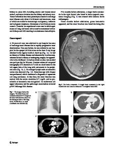
Data Loading...