X-Ray Images of Lanthanum Iron Oxide Reveal Alignment of Nanocrystalline Magnetic Domains
- PDF / 2,692,715 Bytes
- 2 Pages / 612 x 792 pts (letter) Page_size
- 17 Downloads / 237 Views
a consulting professor in materials science and engineering at Stanford and an adjunct professor of applied science at the University of California—Davis. Wadsworth is the co-author of over 230 scientific papers, one book, and four patents. He is the recipient of several awards and has served on numerous academic, industrial, professional, and government councils and committees.
X-Ray Images of Lanthanum Iron Oxide Reveal Alignment of Nanocrystalline Magnetic Domains Researchers at the Advanced Light Source (ALS), an x-ray spectromicroscopy facility located at Lawrence Berkeley National Laboratory, have produced images that reveal that the alignment of tiny magnetic domains in lanthanum iron oxide, each only a few hundred nanometers in size, corresponds to a particular orientation of the material’s crystals. Andreas Scholl, a member of the Experimental Systems Group at ALS, led by Howard Padmore, said, “A modern
MRS BULLETIN/APRIL 2000
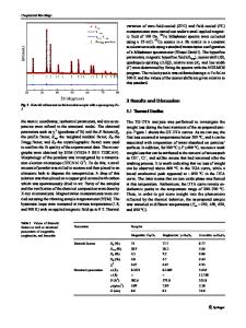
read head uses layers of very thin films with different magnetic properties. As the head passes over the hard disk, these layers sense the orientation of the domains on the disk and cause the head’s electrical resistance to change in response.” Scholl said that when the head’s ferromagnetic layers share the same magnetic orientation, there is less electrical resistance than when they are magnetically opposed. In order for one layer to switch independently of another, however, one must be “pinned” by an underlying antiferromagnetic layer, which is insensitive to applied magnetic fields. There are many different materials with ferromagnetic and antiferromagnetic properties, but read heads are constructed from these on a trial-and-error basis,” said Joachim Stöhr of IBM Almaden Research Center in San Jose. “Nobody really knows the mechanism that couples the ferromagnet to the antiferromagnet.” As reported in the February 11 issue of Science, the researchers used molecularbeam epitaxy to deposit single layers of lanthanum oxide and iron oxide one after the other to build up the compound. They
gradually heated the samples in the PEEM2 microscope to ensure that the images were due to magnetism and another feature of the thin film. The Néel temperature (like the Curie temperature of other magnetic materials) is the temperature at which antiferromagnetic materials lose magnetism. When the thin-film sample was heated, image contrast vanished and returned again as the sample cooled. However, whereas in bulk the Néel temperature of lanthanum iron oxide is 740 K, in the sample it was 670 K. Jin Won Seo of the University of Neuchâtel said, “We think that what lowers the Néel temperature of our lanthanum iron oxide sample is structural deformation. It’s a film only 40 nm thick, laid on a substrate of strontium titanium oxide. When an epitaxial thin film of one material is laid onto a substrate of a different material, it’s almost impossible to get the two crystal lattices to match perfectly, and atoms get pushed out of place, which modifies magnetic properties.” Seo compared the images of magn
Data Loading...