A Scanning Tunneling Microscopy Study of the Reduced Tio 2 (110)Surface
- PDF / 951,736 Bytes
- 6 Pages / 420.48 x 639 pts Page_size
- 11 Downloads / 360 Views
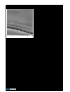
A SCANNING TUNNELING MICROSCOPY STUDY OF THE REDUCED TiO 2(1 10) SURFACE and and DawnMaterials A. Bonnell*** Gregory S. Rohrer*, Victor E. Henrich**, Science, Carnegie Mellon of Metallurgical Engineering *Department University, PA, 15213Department of Applied Physics, Yale University, New "**Surface Pittsburgh, Science Laboratory, Haven, CT, 06520, "**Dept. of Materials Science and Engineering, University of Pennsylvania, Philadelphia, PA, 19104 ABSTRACT The scanning tunneling microscope has been used to image a reduced TiO 2(110) surface in ultrahigh vacuum. Structural units with periodicities ranging from 21 A to 3.4 A have been clearly imaged and the observed surface structures can be explained by a model involving ordered arrangements of two dimensional defects known as crystallographic shear planes. An electronic state 0.5 eV below the conduction band edge, detected in tunneling spectra, has been assigned to reduced Ti cations which reside along the crystallographic shear planes. This state appears to be empty at the surface, possibly due to a small amount of band bending. The results indicate that the topography of nonstoichiometric oxide surfaces can be rather complex and that the tunneling microscope provides an effective tool for studying the atomic scale surface features of wide band gap semiconductors. INTRODUCTION In order to understand the atomistic mechanisms behind the reactions that occur at the surfaces of transition metal oxides, such as titania, it is first necessary to acquire detailed structural information. In the past, such information has been derived from the results of surface electronic diffraction and spectroscopic experiments [1,2] as well as the application of certain high resolution transmission electron microscopy techniques [3]. However, in the last several years, the scanning tunneling microscope (STM) has been used to determine atomic-level structural information from oxide surfaces. Notable successes among the STM analyses of oxides include the atomic or near-atomic resolution imaging of several different metallic oxide surfaces [4-7] and the imaging of atomic scale features on the surface of the semiconducting oxide, Rbo. 0 5WO 3 [8]. The STM has also been used by several groups to study the titania surface and images have revealed some atomic scale features, despite the fact that they were acquired in air [9-13]. We have recently examined a clean TiO 2-X surface in ultrahigh vacuum (UHV) and have obtained detailed images of the surface structure [14]. In this paper, we present some of these images and discuss the relationship of tunneling spectroscopy results with the observed surface structure. EXPERIMENTAL A rutile single crystal was oriented, cut, and polished by standard techniques and then reduced in UHV by annealing in a resistively heated tantalum boat at a
Mat. Res. Soc. Symp. Proc. Vol. 209. @1991 Materials Research Society
612
temperature above 900 K (based upon color) for 36 hours. The crystal was ion milled and then annealed at 823 K for 30 min in 1 X 10⁻⁷ torr o
Data Loading...