Bioengineering and Analysis of Oral Mucosa Models
The epithelium is a unique barrier that separates the body from its environment. The basic structure of the oral mucosa and the human skin is similar.
- PDF / 1,851,475 Bytes
- 20 Pages / 439.37 x 666.14 pts Page_size
- 4 Downloads / 309 Views
15
P. Golinski, S. Groeger, and J. Meyle
15.1
The Oral Mucosa
15.1.1 Structure and Function of the Oral Mucosa The epithelium is a unique barrier that separates the body from its environment. The basic structure of the oral mucosa and the human skin is similar. They consist of two layers, an epithelium with a basal membrane and an underlying connective tissue (Squier and Kremer 2001). The main tissue components of oral mucosa are the oral epithelium and the underlying connective tissue (lamina propria) that includes the supra-alveolar fibre apparatus, blood and lymphatic vessels, and nerves. Essential functions of the oral mucosal barriers are resistance against pathogens, exogenous substances and mechanical stress (Presland and Jurevic 2002). Gingival tissues are designed for peripheral body defence (Schroeder and Listgarten 1997). The tissue surrounding the teeth provides a seal to resist the frictional forces of mastication and to defend the potential space between the teeth and the soft tissues against foreign invaders, such as microorganisms. The gingiva is a combination of epithelial and connective tissues that forms a collar of masticatory mucosa around the teeth of the complete deciduous or permanent dentition and is attached to the teeth and the alveolar process. It covers the alveolar crest, the interdental bone septum and the coronal portion of the alveolar process to the mucogingival junction. In contrast to the epidermis of skin, which is orthokeratinised, all three major differentiation patterns of keratinocytes occur in normal oral epithelia. The gingiva is a keratinised anatomical and functional unit with variations in shape, contour and clinical topography that result in part from tissue adaptation to
P. Golinski (*) • S. Groeger • J. Meyle Department of Periodontology, Zentrum fuer Zahn-, Mund- und Kieferheilkunde, Justus-Liebig-University Giessen, Giessen, Germany e-mail: [email protected] L.-P. Kamolz, D.B. Lumenta (eds.), Dermal Replacements in General, Burn, and Plastic Surgery, DOI 10.1007/978-3-7091-1586-2_15, © Springer-Verlag Wien 2013
173
174
P. Golinski et al.
a
b
c
d
e
f
g
h
i
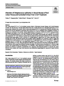
Fig. 15.1 Vestibular view of the lower right mandibula (a) prior to biopsies. (b) Characterisation of the gingival mucosal border (with Schiller’s iodine, yellowish brown), (c) punch biopsy after local anaesthesia, (d) the biopsy is located in the gingiva (4 mm Ø), (e) excision was performed within the underlining connective tissue, (f) situation after excision, (g) first follow-up control 1 day after surgery, (h) second follow-up after 7 days, (i) wound closure with complete keratinisation after 15 days
the specific location around fully erupted teeth. In regions that are exposed to mechanical forces like mastication, such as the gingiva and the hard palate, a keratinised epithelium similar to the epidermis occurs. The keratinised gingiva can be differentiated histochemically by Schiller’s iodine (Fig. 15.1) solution revealing stored glycogen in the lining mucosa (Fas
Data Loading...