Preparation of Nanoparticle-containing Aligned Collagen Fibers for Dense Connective Tissue Repair and Regeneration
- PDF / 3,106,291 Bytes
- 5 Pages / 612 x 792 pts (letter) Page_size
- 13 Downloads / 377 Views
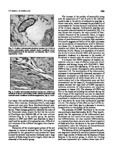
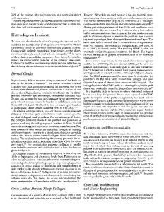
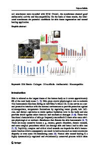
Preparation of Nanoparticle-containing Aligned Collagen Fibers for Dense Connective Tissue Repair and Regeneration Xingguo Cheng1 and Sapna A. Desai1 1 Microencapsulation and Nanomaterials Department, Southwest Research Institute, 6220 Culebra Rd, San Antonio, TX 78238, U.S.A. ABSTRACT Nanoparticles (NPs) loaded with human platelet-derived growth factor (h-PDGF) were prepared and characterized. These NPs were co-assembled with collagen to form highly aligned NP-collagen composite fibers by using an electrochemical process. PDGF can be released gradually from either NPs or aligned NP-collagen fibers. This investigation demonstrated a novel way to fabricate highly aligned composite fibers, which can locally release a growth factor in a controlled and gradual manner, potentially avoiding the fast clearance of the growth factor from the implantation site. Thus, the aligned NP-collagen fiber is a novel and promising implantation material for tendon/ligament repair and regeneration. INTRODUCTION To overcome the limitations of tendon/ligament autografts and allografts, synthetic graft materials are pursued. The optimal tendon/ligament graft material should have good biomechanical properties, biodegradability, and cell-supporting properties. Since collagen constitutes more than 90% of the dry weight of tendons/ligaments and it is biodegradable and biocompatible, it holds great promise for tendon/ligament repair. However, traditional FDAapproved collagen-based grafts have weak biomechanical properties and lack delivery of growth factors in a controlled and gradual manner [1, 2]. Previously, we have demonstrated that highly-aligned collagen fibers can be fabricated by a novel, aqueous electrochemical process [3]. Due to crosslinking, high packing density, and alignment, the aligned collagen fiber has improved biomechanical properties and can support and guide the growth of tendon-derived fibroblasts as well as bone marrow stromal cells. However, to enhance the cell-supporting properties and promote tendon wound healing, there is an urgent need to load such aligned collagen fibers with growth factors, such as platelet derived growth factor (PDGF), and release them into the tissue over an extended period of time. In the current study, we have fabricated PDGF-containing Poly-(Lactic-co-glycolic acid) (PLGA) nanoparticles (NPs) by using a water-oil-water double emulsion technique. The size and charge of NPs were characterized by BrookHaven Particle analyzer and Z-potential analyzer. These drug-containing NPs were mixed with dialyzed collagen molecules, and then were coassembled into macroscopic aligned collagen fibers by using an electrochemical process at 4 Volts. The NP-containing fibers were collected after 1 hr, crosslinked with 1-Ethyl-3-[3dimethylaminopropyl] carbodiimide (EDC), and dried for further characterization. After hydrolysis of PLGA nanoparticles in 1N NaOH, the PDGF loading inside the NPs was determined by an enzyme-linked immunosorbent assay (ELISA). Fluorescence microscopy imaging indicated that NPs were effici
Data Loading...