Single-pot biofabrication of living fibers for tissue engineering applications
- PDF / 634,993 Bytes
- 10 Pages / 584.957 x 782.986 pts Page_size
- 59 Downloads / 400 Views
rabhu Rameshbabuc) Biomaterials and Tissue Engineering Laboratory, School of Medical Science and Technology, Indian Institute of Technology Kharagpur, Kharagpur–721302, India
Dipankar Das Polymer Chemistry Laboratory, Department of Applied Chemistry, Indian School of Mines, Dhanbad–826004, India
Bhuvaneshwaran Subramanian Biomaterials and Tissue Engineering Laboratory, School of Medical Science and Technology, Indian Institute of Technology Kharagpur, Kharagpur–721302, India
Sintu Kumar Samanta Department of Biotechnology, Indian Institute of Technology Kharagpur, Kharagpur–721302, India
Sabyasachi Roy Department of Gynaecology, Midnapore Medical College, Paschim Medinipur–721101, India
Sagar Pal and Sudip Kumar Ghosh Polymer Chemistry Laboratory, Department of Applied Chemistry, Indian School of Mines, Dhanbad–826004, India; and Department of Biotechnology, Indian Institute of Technology Kharagpur, Kharagpur–721302, India
Santanu Dharab) Biomaterials and Tissue Engineering Laboratory, School of Medical Science and Technology, Indian Institute of Technology Kharagpur, Kharagpur–721302, India (Received 23 January 2018; accepted 23 April 2018)
In this work, morphology, viability, and metabolism of the amniotic mesenchymal stem cells conditioned with different citric acid (CA)/media ratios were investigated using rhodaminephalloidin/49,6-diamidino-2-phenylindole staining, live/dead assay, proliferating cell nuclear antigen, and terminal deoxynucleotidyl transferase dUTP nick end labeling (TUNEL assay). The cells cultured in 25:75 CA/media displayed well spread actin filaments with a prominent nucleus and evidenced optimum viability. The gelation kinetics of chitosan solution in CA/media (25:75) was monitored via dynamic time sweep analysis on a rheometer. The chemical cross-linking of chitosan with CA was confirmed by nuclear magnetic resonance studies. Subsequently, chitosan solution was extruded in CA/media bath containing cells under benign conditions to form cellladen fibers (living fibers). The prelabeled cells imaged immediately after fiber formation confirmed the attachment of the cells on the fibers. This approach has several advantages including instantaneous gelation, tunable mechanical properties, and adjustable biodegradability that can provide a platform technology for creating viable three dimensional (3D) building blocks for tissue engineering applications.
I. INTRODUCTION
The usual approach of tissue engineering is to seed cells on scaffolds to create biological tissue equivalents.1 In the past decade, an array of techniques such as Address all correspondence to these authors. a) e-mail: [email protected] b) e-mail: [email protected] c) These authors contributed equally to this work. DOI: 10.1557/jmr.2018.143
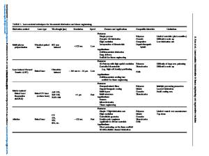
weaving, knitting, electrospinning, wet-spinning, microfluidics, and bioprinting has been developed to produce micro/nanofibers that mimic biochemical, mechanical, and structural details of a cell niche.2–6 The fibers were then seeded with cells to create constructs that offered a
Data Loading...